Search This Blog

Friday, October 7, 2022

Promising medication for sleep apnea

 Targeting a condition suffered by nearly a billion people worldwide, a new study from Flinders University has shown a drug previously used to treat depression can reduce obstructive sleep apnea (OSA) severity.

While not yet identifying a cure, the authors say the study opens up further avenues for the development of future  treatments targeted at the huge number of people unable to tolerate current sleep apnea therapies, such as  (CPAP) machines.

"Obstructive sleep apnea can be a debilitating disease, causing  at night and sleepiness during the day," says study lead author Dr. Thomas Altree from FHRMI: Sleep Health (formerly the Adelaide Institute for Sleep Health).

"It affects millions of Australians and causes major impacts on health and productivity."

"Recent research found a combination of the medicines reboxetine and oxybutynin, which were both previously used for unrelated conditions, could be an  for obstructive sleep apnea but can cause side effects."

"We wanted to see if reboxetine on its own could be effective and assess exactly how it changes breathing during sleep."

The team ran a double blind, placebo controlled, randomized, multicenter cross-over trial with collaborators at the Woolcock Institute in Sydney (following a gold standard for drug trials) with 16 people who had OSA. They tested single doses of reboxetine compared to a combination of reboxetine and oxybutynin or placebo.

"Our results showed that reboxetine on its own can reduce sleep apnea severity," says Dr. Altree.

"We found the drug reduced the number of sleep apnea events per hour and also improved , while the addition of oxybutynin didn't cause additional improvements."

"We also used a state-of-the-art computing method to determine how the drug stabilizes breathing during sleep, which allows us to identify which patients might benefit most from the drug in the future."

The team's findings present the first evidence that reboxetine alone reduces OSA severity, and provides further insight into the role of norepinephrine reuptake inhibitors on upper airway stability during sleep.

"The current gold-standard treatment of sleep apnea is with a CPAP device during sleep. But this one-size-fits-all approach doesn't address the fact that there are different causes for sleep apnea. In addition, many people can't tolerate CPAP in the long term," says Dr. Altree.

"It's therefore important we discover other avenues to assist people, and this study provides an important step for future drug development."

The paper is published in the Journal of Clinical Sleep Medicine.


Explore further
Rehospitalization is lower in adults with heart disease when they are treated for comorbid sleep apnea

More information: Thomas J. Altree et al, The norepinephrine reuptake inhibitor reboxetine alone reduces obstructive sleep apnea severity: a double blind, placebo controlled, randomized, cross-over trial, Journal of Clinical Sleep Medicine (2022). DOI: 10.5664/jcsm.10256
https://medicalxpress.com/news/2022-10-team-medication-apnea.html

Discovery of additional function of cerebellum

 The cerebellum is known primarily for regulation of movement. Researchers at the University of Basel have now discovered that the cerebellum also plays an important role in remembering emotional experiences. The study appears in the journal PNAS.

Both positive and  are stored particularly well in memory. This phenomenon is important to our survival, since we need to remember  in order to avoid them in the future. Previous studies have shown that a  called the amygdala, which is important in the processing of emotions, plays a central role in this phenomenon. Emotions activate the amygdala, which in turn facilitates the storage of information in various areas of the cerebrum.

The current research, led by Professor Dominique de Quervain and Professor Andreas Papassotiropoulos at the University of Basel, investigates the role of the cerebellum in storing . In a large-scale study, the researchers showed 1,418 participants emotional and neutral images and recorded the subjects' brain activity using magnetic resonance imaging.

In a memory test conducted later, the positive and negative images were remembered by the participants much better than the neutral images. The improved storage of emotional images was linked with an increase in  in the areas of the cerebrum already known to play a part. However, the team also identified increased activity in the cerebellum.

The cerebellum in communication with the cerebrum

The researchers were also able to demonstrate that the cerebellum shows stronger communication with various areas of the cerebrum during the process of enhanced storage of the emotional images. It receives information from the cingulate gyrus—a region of the brain that is important in the perception and evaluation of feelings. Furthermore, the cerebellum sends out signals to various regions of the brain, including the amygdala and hippocampus. The latter plays a central role in memory storage.

"These results indicate that the  is an integral component of a network that is responsible for the improved storage of emotional information," says de Quervain. Although an improved memory for emotional events is a crucial mechanism for survival, it does have its downsides: in the case of very negative experiences, it can lead to recurring anxiety. This means that the findings, which have now been released, may also be relevant in understanding psychiatric conditions such as post-traumatic stress disorder.


Explore further

Why do we remember stressful experiences better?

More information: Matthias Fastenrath et al, Human cerebellum and corticocerebellar connections involved in emotional memory enhancement, Proceedings of the National Academy of Sciences (2022). DOI: 10.1073/pnas.2204900119
https://medicalxpress.com/news/2022-10-discovery-function-cerebellum.html

Detecting Alzheimer's disease in the blood

 Researchers from Hokkaido University and Toppan have developed a method to detect build-up of amyloid β in the brain, a characteristic of Alzheimer's disease, from biomarkers in blood samples.

Alzheimer's disease is a neurodegenerative disease, characterized by a gradual loss of neurons and synapses in the . One of the primary causes of Alzheimer's disease is the accumulation of amyloid β (Aβ) in the brain, where it forms plaques. Alzheimer's disease is mostly seen in individuals over 65 years of age, and cannot currently be stopped or reversed. Thus, Alzheimer's disease is a major concern for nations with aging populations, such as Japan.

A team of scientists from Hokkaido University and Toppan, led by Specially Appointed Associate Professor Kohei Yuyama at the Faculty of Advanced Life Science, Hokkaido University, have developed a biosensing technology that can detect Aβ-binding exosomes in the blood of mice, which increase as Aβ accumulates in the brain. Their research was published in the journal Alzheimer's Research & Therapy.

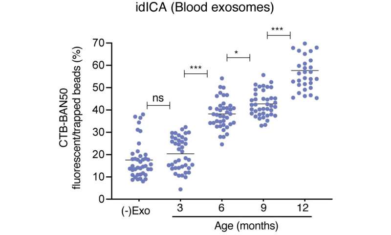
When tested on mice models, the Aβ-binding  Digital ICA (idICA) showed that the concentration of Aβ-binding exosomes increased with the increase in age of the mice. This is significant as the mice used were Alzheimer's disease model mice, where Aβ builds up in the brain with age.

In addition to the lack of effective treatments of Alzheimer's, there are few methods to diagnose Alzheimer's. Alzheimer's can only be definitively diagnosed by direct examination of the brain—which can only be done after death. Aβ accumulation in the brain can be measured by cerebrospinal fluid testing or by ; however, the former is an extremely invasive test that cannot be repeated, and the latter is quite expensive. Thus, there is a need for a diagnostic test that is economical, accurate and widely available.

Previous work by Yuyama's group has shown that Aβ build-up in the brain is associated with Aβ-binding exosomes secreted from neurons, which degrade and transport Aβ to the microglial cells of the brain. Exosomes are membrane-enclosed sacs secreted by cells that possess cell markers on their surface. The team adapted Toppan's proprietary Digital Invasive Cleavage Assay (Digital ICA) to quantify the concentration of Aβ-binding exosomes in as little as 100 µL of blood. The device they developed traps molecules and particles in a sample one-by-one in a million micrometer-sized microscopic wells on a measurement chip and detects the presence or absence of fluorescent signals emitted by the cleaving of the Aβ-binding exosomes.

Detecting Alzheimer's disease in the blood
Concentration of amyloid β-binding exosomes that were detected by the Digital ICA chip in the blood of mice of different ages. asterisks represent significant results. Credit: Kohei Yuyama, et al. Alzheimer's Research & Therapy. October 3, 2022

Clinical trials of the technology are currently underway in humans. This highly sensitive idICA technology is the first application of ICA that enables highly sensitive detection of exosomes that retain specific surface molecules from a small amount of blood without the need to learn special techniques; as it is applicable to exosome biomarkers in general, it can also be adapted for use in the diagnosis of other diseases.


Explore further

New mechanism by which Alzheimer's disease spreads through the brain discovered

More information: Kohei Yuyama et al, Immuno-digital invasive cleavage assay for analyzing Alzheimer's amyloid ß-bound extracellular vesicles, Alzheimer's Research & Therapy (2022). DOI: 10.1186/s13195-022-01073-w
https://medicalxpress.com/news/2022-10-alzheimer-disease-blood.html

ALS patients' own cells may provide a safe pathway to slow or halt progression

 Removing ALS (amyotrophic lateral sclerosis) patients' own dysfunctional cells, fixing them, then putting them back in patients' bodies is a safe, well tolerated process that has been shown to slow or halt disease progression in a small number of patients, according to a study by the Houston Methodist Research Institute and Massachusetts General Hospital.

The study, published recently in the journal Neurology: Neuroimmunology and NeuroInflammation, was designed as a randomized, placebo-controlled, phase 2a trial with 12 participants followed by an open-label trial. But the unpredictability of the COVID-19 pandemic—which interrupted and even halted many research projects around the country—led to a reduction of the enrollment to 7 participants in the double-blind trial and 8 participants in the six-month open label trial.

Seven ALS patients completed the Group 1 randomized placebo-controlled trial, while eight participated in the Group 2 open label portion of the study. Both groups had dysfunctional regulatory T-cells—or Tregs—withdrawn from their bodies. When functioning normally, Tregs help reduce inflammation. In all but the placebo control group, researchers expanded the faulty Tregs to restore their function, then infused them back into patients' bodies, along with a low dose of Interleukin-2 to help boost the immune system.

Results showed the Treg treatments were 100% safe and tolerable, and they remained biologically active in ALS patients for more than a year. The treatments also slowed or halted disease progression in most participants.

Participants included Houston Methodist patients Darci Garcia, enrolled in Group 1, and John Lay, from Group 2, both of whom reported a slowdown of ALS progression during and for a short time after the clinical trial treatments.

"We look forward to a much larger clinical trial that will allow for proper evaluation of clinical efficacy and further characterization of the long-term safety of this therapy," said Dr. Jason Thonhoff, principal investigator. "These early results in a handful of patients are promising."

The open label segment of this latest Treg trial led to a spinoff publication reporting the identification of two biomarkers that could potentially help track ALS  and measure just how well ALS therapies work.

More information: Jason R. Thonhoff et al, Combined Regulatory T-Lymphocyte and IL-2 Treatment Is Safe, Tolerable, and Biologically Active for 1 Year in Persons With Amyotrophic Lateral Sclerosis, Neurology—Neuroimmunology Neuroinflammation (2022). DOI: 10.1212/NXI.0000000000200019

https://medicalxpress.com/news/2022-10-als-patients-cells-safe-pathway.html

Protein that potentially worsens kidney injuries IDd

 One of the many risks associated with heart attacks and cardiac surgery is acute kidney injury, a serious condition that in some cases can lead to kidney failure.

Common as it is—it's observed 10% to 20% of the time following  or a — scientists don't have a full biological understanding of why the  is happening.

"There are some conventional explanations, but I felt those were not fully accurate," said Dr. Rajesh Gupta, an associate professor of medicine in The University of Toledo College of Medicine and Life Sciences. "I'm challenging the conventional explanations to say there are additional mechanisms at play, and we need to understand this in a detailed way."

A new study from UToledo published in the journal Kidney International shows that the accumulation of a protein called hemopexin in the kidney is partly to blame—a finding that could have implications in a range of kidney injuries.

Gupta, who works as an interventional cardiologist at The University of Toledo Medical Center, had taken note of a decade-old study that found hemopexin was accumulating in damaged kidneys.

Part of the body's natural defense system, hemopexin bonds to and clears excess heme from the blood. While heme is essential for handling and delivering oxygen within our cells, it can be toxic when too much has been freely released into the bloodstream—something that occurs following many acute illnesses.

It was an interesting observation but no one had yet identified what hemopexin was doing in the kidney.

"Is this good or is it bad? Hemopexin is normally considered a protective protein. Maybe it's doing something beneficial," Gupta said. "Or you could hypothesize that it's normally protective, but in this case, it's stuck in the kidney and part of the problem. We just didn't know."

UToledo researchers sought to answer that question by using a pair of mouse models. One model was normal, while the other had been genetically altered so it could not produce hemopexin. The two groups were then studied in models of acute kidney injury to determine the role of hemopexin.

The team found important differences between the two groups.

Following an acute kidney injury, mice that naturally produced hemopexin experienced significantly worse kidney damage and lower renal function than the mice that could not produce the protein.

The team also found higher levels of a key indicator of oxidative stress in the hemopexin-producing mice.

"Hemopexin has a surprisingly detrimental effect on kidney injury. Essentially, it's getting stuck in the kidney and it's bringing with it hemoglobin, which mediates an iron toxicity injury," Gupta said. "Although iron has important normal functions in oxygen handling, it can be problematic if it's in the wrong place under the wrong circumstances. Iron can be like a firecracker or explosive, creating all these interactions that are inflammatory."

Acute kidney injuries are common, particularly in patients who are seriously ill and hospitalized. As the kidneys rapidly lose their ability to filter waste from the body, harmful substances can build up in the blood, causing damage to other organs.

Without prompt treatment even relatively minor acute kidney injuries can lead to lasting damage. Generally, treatment involves addressing the underlying issue that caused the injury.

"Because acute kidney injuries have a range of causes, there is no . The treatment varies based on the specific cause and typically involves supportive care. If it turns out that hemopexin is playing a role, regardless of what brought on the injury, that could be a major turning point in our understanding of kidney injury," said Dr. Xiaoming Fan, a postdoctoral fellow in the College of Medicine and Life Sciences and the paper's first author.

With the knowledge that hemopexin is intensifying renal damage following an , it could provide a more targeted therapy—if doctors could prevent hemopexin from accumulating in the kidney, they might be able to lessen the damage.

The finding also raises concerns about an emerging interest in hemopexin as a therapy of its own. The protein is being investigated for the potential to treat , sepsis and complications arising from extensive blood transfusions.

While laboratory experiments have shown promise in hemopexin's ability to lessen inflammation and tissue damage in those conditions, Gupta said it's important that researchers now consider a previously unknown side effect.

"Our findings say we should carefully look at kidney health if we're potentially testing hemopexin as a therapeutic," he said.


Explore further

Acute kidney injury among African Americans with sickle cell trait and disease

More information: Xiaoming Fan et al, Hemopexin accumulates in kidneys and worsens acute kidney injury by causing hemoglobin deposition and exacerbation of iron toxicity in proximal tubules, Kidney International (2022). DOI: 10.1016/j.kint.2022.07.024
https://medicalxpress.com/news/2022-10-protein-potentially-worsens-kidney-injuries.html

Ultrasound can move, reposition or break up kidney stones while patient is awake

 A new technique which combines the use of two ultrasound technologies may offer an option to move kidney stones out of the ureter with minimal pain and no anesthesia, according to a new feasibility study published today in the Journal of Urology.

In the procedure, the physician uses a handheld transducer placed on the skin to direct ultrasound waves towards the . The ultrasound can then be used to move and reposition the stones to promote their passage, a process called ultrasound propulsion, or the break up the stone, a technique called burst wave lithotripsy (BWL).

Unlike shock wave lithotripsy, which is the standard procedure now in use and requires sedation, this technology doesn't hurt, said lead author Dr. M. Kennedy Hall, a UW Medicine emergency medicine doctor. "It's nearly painless, and you can do it while the patient is awake, and without sedation, which is critical."

The research team hopes that, with this new technology, the procedure of moving or breaking up the stones could eventually be performed in a clinic or emergency room setting, Hall added.

Stones in the ureter, which leads from the kidney to the bladder, can cause  and are a common reason for emergency department visits. Most patients with ureteral stones are advised to wait to see if the stone will pass on its own. However, this observation period can last for weeks, with nearly one-fourth of patients eventually requiring surgery, Hall noted.

One in 11 Americans will have a urinary stone over the course of their lifetime. The incidence appears to be increasing, according to one UW Medicine study looking at this same technology. Up to 50% of patients with a stone event will recur within five years, the study noted.

Hall and colleagues evaluated the new technique to meet the need for a way to treat stones without surgery.

The study was designed to test the feasibility of using the ultrasonic propulsion or using BWL to break up stones in awake, unanaesthetized patients, Hall said.

Twenty-nine patients participated in the study. Sixteen were treated with propulsion alone and 13 with propulsion and burst wave lithotripsy. In 19 patients, the stones moved. In two cases, the stones moved out of the ureter and into the bladder.

Burst wave lithotripsy fragmented the stones in seven of the cases. At a two-week follow up, 18 of 21 patients (86%) whose stones were located lower in the ureter, closer to the bladder, had passed their stones. In this group, the average time to stone passage was about four days, the study noted.

One of these patients felt "immediate relief" when the stone was dislodged from the ureter, the study stated.

The next step for the researchers would be to perform a clinical trial with a , which would not receive either BWL bursts or ultrasound propulsion, to evaluate the degree to which this new technology potentially aids stone passage, Hall said.

Development of this technology first started five years ago, when NASA supported a study to see if kidney stones could be moved or broken up, without anesthesia, on long space flights, such as the Mars missions. The technology has worked so well that NASA has downgraded  as a key concern.

"We now have a potential solution for that problem," Hall said.


Explore further

First human study using sound waves to break up kidney stones shows promising results

More information: M. Kennedy Hall et al, First Series Using Ultrasonic Propulsion and Burst Wave Lithotripsy to Treat Ureteral Stones, Journal of Urology (2022). DOI: 10.1097/JU.0000000000002864
https://medicalxpress.com/news/2022-10-ultrasound-reposition-kidney-stones-patient.html

Fast track to fertility program halves time to treatment

 When struggling to conceive, every second that ticks by feels precious. That makes it easy to get discouraged: 65 percent of those who seek fertility care eventually discontinue treatment, the majority due to stress. That's why Penn Medicine recently instituted a telemedicine-driven program aimed at seeing patients more quickly and starting treatments sooner. The program, Fast Track to Fertility, cut the time between when patients initially reached out for help to when they received their first treatment by half—getting them on the path to parenthood roughly a month and a half sooner, according to research published in NEJM Catalyst by researchers at the Perelman School of Medicine at the University of Pennsylvania.

On top of cutting the average time it took for new patients to get their first treatment (from 97 to 41 days), the program also allowed for more new patients to access fertility care, increasing the number by 24 percent in the year it was implemented as a standard of care at Penn Medicine. At a time when one in eight couples in the United States are experiencing infertility, Fast Track to Fertility allowed more than 1,000 new patients to begin treatments to help them become pregnant.

"Most of the people who seek fertility care have been trying to get pregnant for at least a year, so the emotional stakes are high and they really want to get started as soon as possible," said the study's senior author and Fast Track to Fertility co-founder, Anuja Dokras, MD, Ph.D., a professor of Obstetrics and Gynecology and chair of Gynecology for the Women's Health Service Line. "Our findings show that this program can significantly speed up the time to treatment and, in so doing, opens the door for so many more people. These findings show this way of doings things can make real differences in people's lives."

Fertility care has steadily increased in demand since it was introduced, reaching a point where fertility clinics often have long waits for new patients. Fast Track to Fertility, started through the Innovation Accelerator in Penn Medicine's Center for Health Care Innovation, seeks to speed things up by deploying a relatively small team of advanced practice providers for efficient telemedicine-based initial visits with new patients as quickly as possible. That visit also gives patients the opportunity to enroll in an artificial intelligence-guided text messaging program that helps guide them through the complex fertility "workup" swiftly and with as few hiccups as possible.

"This system has made it so that as soon as a patient contacts us, their journey begins," said the study's lead author and Fast Track to Fertility co-founder, Suneeta Senapati, MD, an assistant professor of Obstetrics and Gynecology. "Both partners in a couple need to complete a workup, which can include blood work, ultrasounds, X-rays, semen analysis, and more. Some parts of this are dependent on the , making it a time-sensitive process, so making it as easy as possible to quickly work through—with minimal confusion—is invaluable."

Initial pilots (which used human texters instead of  to test the system) reduced the wait time to new patient visits from initial contact with the practice by 88 percent, making the average wait time just four days. And no patients during those first pilots had to call the office to figure out next steps, compared to a quarter of patients who weren't in the program.

In 2021, when the latest analysis was performed, Fast Track to Fertility was expanded to become the standard of care across Penn Medicine's department of Obstetrics and Gynecology. In addition to halving the time to treatment and increasing new patients, they also saw appointment "no-shows"—which includes those who unexpectedly don't go to their appointment or who need to cancel late—drop from 40 to 20 percent, a particularly important measure.

"Any time there is a no-show, we can't backfill that appointment because of the  that goes into this type of care," Dokras said. "So any time we can reduce no-shows, that means more people can get the care they're looking for to start their family."

Satisfaction, both from the patients and the advanced practice providers running the system, was also found to be high. And the researchers hope that the system can be expanded to support patients throughout their fertility journey.

"These care models do not replace our clinical work force, as  remains imperative to the doctor-patient relationship and care delivery. Rather, they improve efficiency—while maintaining personalized care—for both  and their care teams to accommodate the growing demands for fertility services," Senapati said. "In the end, this enables my colleagues and I to do more of what we got into this field for: Helping people get pregnant and bring home their babies."


Explore further

Continued access to fertility preservation is critical for teens and young adult cancer patients after SCOTUS decision

More information: Suneeta Senapati et al, The Fast Track to Fertility Program: Rapid Cycle Innovation to Redesign Fertility Care, NEJM Catalyst (2022). DOI: 10.1056/CAT.22.0065
https://medicalxpress.com/news/2022-10-fast-track-fertility-halves-treatment.html