Search This Blog

Saturday, August 13, 2022

The New Polio Outbreak Is Bad. But Why Hasn't It Become a Disaster (Yet)?

 Viruses are fascinating. Watching the twists and turns of SARS-CoV-2 mutations and their effect on the entire planet has made it easy to appreciate the raw evolutionary ability of a novel viral pathogen. But polio… there’s a virus with some serious history on its side, from Egyptian carvings to FDR’s wheelchair, and it’s still grabbing headlines.

The latest:

It’s easy to feel a bit of panic in the air. A young man was paralyzed in the New York City suburbs. Poliovirus was found in the wastewater of 8 boroughs of London. What in Sam Hill is going on here?

To understand, I had to do some reading. Whatever little bit I learned about polio in med school I promptly forgot, since, well, no one sees polio anymore. Vaccines made the worst-case, 1 in 200, outcome of a poliovirus infection, paralytic poliomyelitis, virtually disappear. Not totally, however, and that’s where this virus and the vaccines that prevent it get so interesting. So much so that it’s worth breaking it down a little, to get a better handle on why it popped up in a New York City suburb, and what that means to us going forward.

Interesting Polio Vaccine Fact #1The Salk vaccine, generally known as the inactivated polio vaccine (IPV), developed first and used exclusively in the US since 2000, is incredibly effective (99%+) at preventing paralytic poliomyelitis but does not convey mucosal immunity, and therefore does little to reduce transmission of the poliovirus.

Yes, a true life-saving, disability-sparing vaccine, and one with virtually no adverse effects. However, the inactivated virus triggers a marvelous immune response in the blood without establishing immunity in the gut. Since poliovirus is a fecal-oral route pathogen — meaning viral particles are not passed in tiny aerosols from our breath, or large droplets from a cough, but rather in tiny bits of fecal matter that make their way onto hands, or into the water supply, and then into mouths — the poliovirus can readily spread itself in its usual manner among a population entirely immunized via the IPV vaccine. Remember that, Americans! Lest we think our adult selves too clean for such a lowly route of transmission, recall that we are a “wipe right/shake right” society, and that not every food preparer employs ideal hand washing technique in the restaurant bathroom.

When you see Science reporting like this, you might want to unsubscribe; this quite misconstrues the protection actually afforded to a country well-vaccinated via IPV:

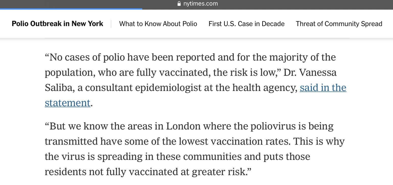
The New York Times offered a similarly poor characterization; it’s fair to fault communities with low IPV uptake for putting their populace at risk, but not for spreading the disease:

Interesting Polio Vaccine Fact #2The live attenuated Oral Polio Vaccine, developed by Dr. Albert Sabin, is cheap, easy (taken as a few drops of liquid by mouth), and remarkably effective both at preventing infection in the gut as well as severe neurological disease, but has an Achilles heel: it can mutate back into a virulent form of polio!

This is the vaccine that has virtually eliminated wild-type polio from the globe. In the US, it was used exclusively for decades until polio was essentially gone, and then IPV reintroduced. Why bring back the older, more expensive IPV? The faint risk of vaccine-associated paralytic polio (VAPP). Very rarely — in the 1 in 2-3 million range — people who receive the vaccine, often immune-compromised, brew up a case of paralyzing poliomyelitis if the virus mutates into a pathogenic form and attacks the nervous system. Sometimes, too, especially in communities employing the OPV vaccine but with lower rates of acceptance, a strain could circulate among the community and acquire mutations over a year or two that would revert it back to a pathogenic strain, so-called “circulating vaccine-derived poliovirus” (cVDPV). The treatment, at least until 2020, was to give everybody more OPV until herd immunity is built up and hope to avoid more vaccine-associated or vaccine-derived cases of polio!

Interesting Polio Vaccine Fact #3: There are 3 types of wild poliovirus; type 2 has been eradicated (and recently type 3 as well) through the global OPV campaign, but type 2 most easily mutates back into a virulent form from the OPV vaccine, meaning that no wild type 2 poliovirus is now in existence, but most cases of actual poliomyelitis are caused by vaccine-derived type 2 polioviruses.

This has led to a wild cat-and-mouse game between vaccine-derived virus and vaccine. Bivalent (type 1 and 3 only) OPV was developed, since wild type 2 was eliminated from the globe and the type 2 strain in the vaccine was so problematic. Great — except when a vaccine-derived type 2 strain begins to circulate in an undervaccinated community! Neither existing option was appealing: either bring in IPV (not always possible in the developing world) only to prevent paralytic disease without breaking transmission chains, or saturate the community with the old trivalent OPV vaccine and risk more of the same problem in the future. The solution to this cycle has been the development of a modified type 2 monovalent OPV (mOPV2), reformulated to greatly reduce the risk of mutating into a problematic form. This is the new band-aid for outbreaks of vaccine-derived poliovirus since 2020; but of course will not protect against the type 1 wild poliovirus still found in Afghanistan and Pakistan, nor the type 3 vaccine-derived poliovirus found circulating in Israel that led to paralysis in a 3 year-old earlier this year.

Speaking of Israel, it appears to be a poorly-kept secret (since the New York Times reported it) that the young man now paralyzed with poliomyelitis from a type 2 vaccine-derived poliovirus is a member of the Orthodox Jewish community. Yes, that Orthodox Jewish community in Rockland and Orange Counties, with the notoriously low rate of childhood vaccinations (around 60%) that went through a measles outbreak in 2018-19.

If we were in Africa or India, the sensible public health response would have been to bring mOPV2 into the community health centers, and have the rabbis begging their synagogues to have everyone immunized with this ideal vaccine to both stop the spread of this strain and prevent severe disease. Instead, in a move that appears to leave virologists somewhere on the spectrum between bemused and bedeviled, we stick with IPV, allowing transmission to continue almost unabated. But at least we publicize it!

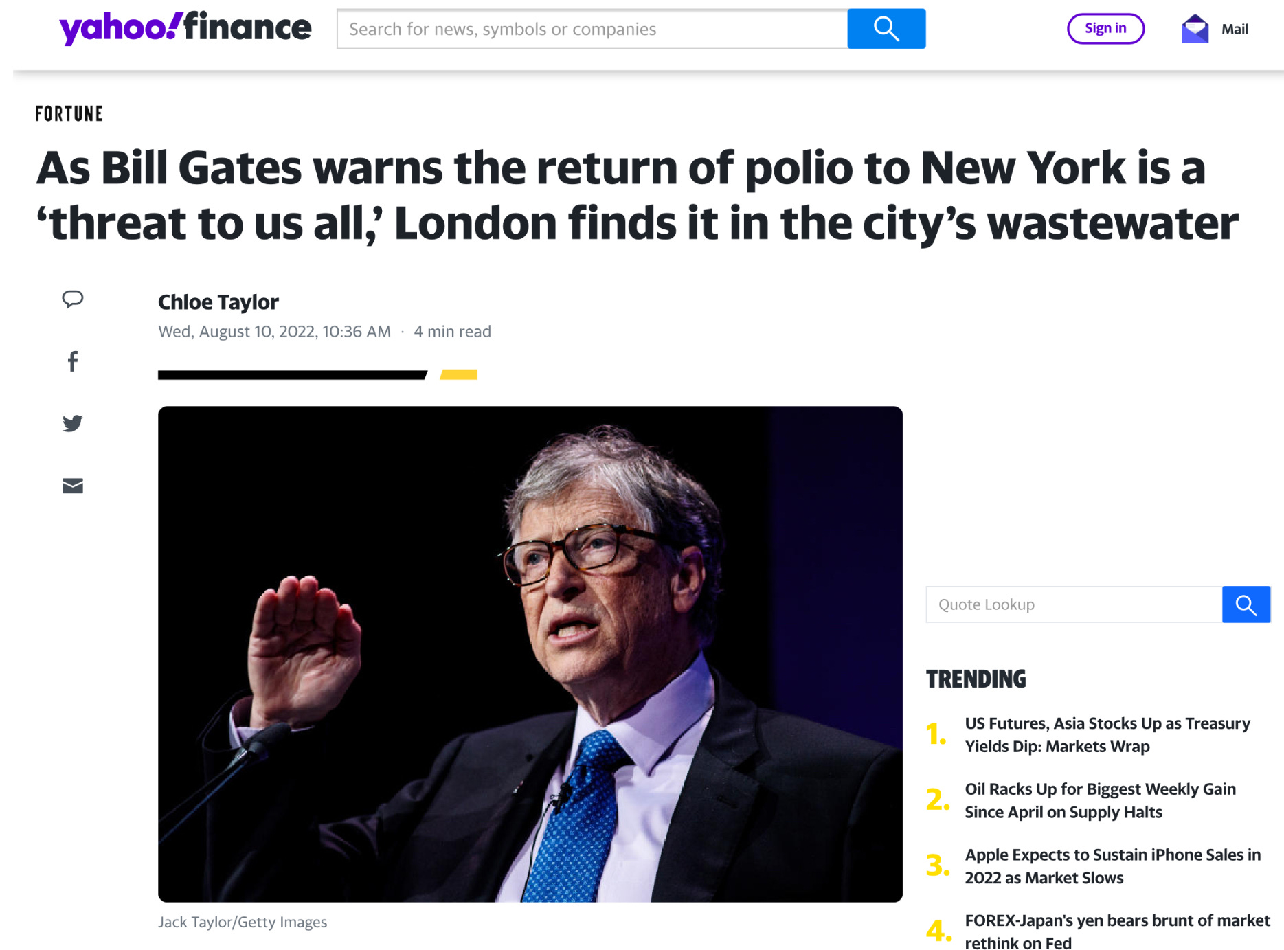
The problem, of course, is that while paralysis will be prevented in people, like the gentleman pictured above, who get their IPV vaccine, there can be no herd immunity via these vaccines for those who can’t be persuaded to roll up their sleeves, given the lack of mucosal immunity. From what I can tell, this creates a problem; observe the reproductive number of polio in a non-immune community:

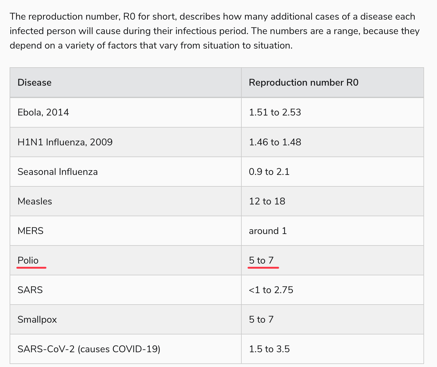
Yes, polio is right up there with our most transmissible pathogens, probably not far from our new friend, Omicron BA.5. Perhaps this is outdated information, calculated in an era of open sewers, or simply overstated. But hearing virologists talk about polio, it’s generally with a sense of, “oh, everyone just gets it” in outbreak conditions, which is why a paralytic polio rate of 1 in 200, or even 1 in 1000, is so problematic.

That, to me, is the mystery of these recent vaccine-derived polio outbreaks. The protection against polio infection — not paralytic poliomyelitis, mind you, just the majority of poliovirus infections which are asymptomatic (around 70-75%) or the remainder that are more flu-like — should be incredibly low, at least among those born after 2000 in the U.S. who are not blessed with the superior immunity of OPV recipients. How does a virus with an R0 >5 circulate for months in places like New York and London without tearing through the kids in a densely-packed, non-immune community, and not rapidly spread to other communities?

https://doctorbuzz.substack.com/p/the-new-polio-outbreak-is-bad-but

New clue may explain who has long COVID—and how to treat

 Public health officials are scrambling to understand long COVID, the condition in which patients report symptoms like fatigue, muscle weakness, and “brain fog” months after infection. A new study released Wednesday shows one way doctors might diagnose who has the chronic condition and indicates a possible way to treat it.

Researchers from the Yale School of Medicine and the Icahn School of Medicine at Mount Sinai found that long COVID patients had half the levels of cortisol—a hormone that guides the body’s response to stress—as uninfected individuals and those who had recovered fully from COVID. The study, which has yet to be peer reviewed, analyzed 215 individuals, 99 of whom were long COVID patients.

If the study’s findings are corroborated, low cortisol levels could help doctors determine who has long COVID, allowing for better treatment. It might also help public health officials better understand how widespread the chronic condition is as long COVID threatens to pull millions of workers from the U.S. labor force.

Scientists are still trying to understand the myriad ways COVID affects the human body. In addition to fatigue and muscle weakness, COVID has also been connected to higher rates of depression, hair losscardiovascular disease, and heart attacks. An earlier study found that even a mild case of COVID led to a decline in brain tissue equivalent to a decade’s worth of aging. And last year, a study found that COVID attacked fat cells, perhaps explaining why overweight and obese individuals are more at risk for severe COVID.


Cortisol is a hormone that helps regulate bodily functions like blood pressure, digestion, and sleep cycles as a person experiences stress. Amid stress, cortisol levels rise, encouraging the body to increase the brain’s use of glucose and release substances that repair tissue, while suppressing bodily functions that are nonessential in a dangerous situation.

Having less of a stress hormone might sound like a good thing, but abnormally low levels of cortisol have been connected to symptoms like muscle weakness, persistent fatigue, a loss of appetite, and low blood pressure. 

Researchers have reported low cortisol levels in those with chronic fatigue syndrome, another long-lasting medical condition whose causes and symptoms have puzzled scientists. Studies have found that low-dose cortisol treatments have helped patients with chronic fatigue. 


Governments are quickly realizing that long COVID presents a threat to the economy. The U.S. government is spending $1.2 billion to determine the causes behind the condition and research possible treatments.

The U.S. Centers for Disease Control and Prevention estimates that one in five American adult COVID-19 patients is suffering from symptoms associated with long COVID. Katie Bach, a senior fellow at the Brookings Institution and an expert on long COVID’s effect on the workforce, estimates that up to 4 million workers, or 2.4% of the U.S. labor force, is out of the workforce as a result of long COVID. Other economies are reporting even higher rates of the condition, with the U.K. reporting in April that long COVID was hurting 4% of the country’s workforce. 

Wednesday’s study may help doctors treat long COVID. Akiko Iwasaki, one of the study’s authors, told Bloomberg that the research provides “many clues for therapeutic avenues, including antivirals and hormone therapy.”

https://fortune.com/2022/08/12/long-covid-causes-treatment-stress-hormone-cortisol-study/

Circulating protein associated with progression to end-stage kidney disease in diabetes

 HIROKI KOBAYASHI HTTPS://ORCID.ORG/0000-0002-9674-0374HELEN C. LOOKEREIICHIRO SATAKE HTTPS://ORCID.ORG/0000-0002-9784-4423FRANCESCA D’ADDIO HTTPS://ORCID.ORG/0000-0002-0345-0694JONATHAN M. WILSON HTTPS://ORCID.ORG/0000-0002-8648-600XPIERRE JEAN SAULNIER HTTPS://ORCID.ORG/0000-0003-1862-4252ZAIPUL I. MD DOM HTTPS://ORCID.ORG/0000-0003-3503-7391KRISTINA O’NEIL HTTPS://ORCID.ORG/0000-0002-2020-8728KATSUHITO IHARA HTTPS://ORCID.ORG/0000-0001-9554-3646[...]ANDRZEJ S. KROLEWSK

DOI: 10.1126/scitranslmed.abj2109

Abstract

Circulating proteins associated with transforming growth factor–β (TGF-β) signaling are implicated in the development of diabetic kidney disease (DKD). It remains to be comprehensively examined which of these proteins are involved in the pathogenesis of DKD and its progression to end-stage kidney disease (ESKD) in humans. Using the SOMAscan proteomic platform, we measured concentrations of 25 TGF-β signaling family proteins in four different cohorts composed in total of 754 Caucasian or Pima Indian individuals with type 1 or type 2 diabetes. Of these 25 circulating proteins, we identified neuroblastoma suppressor of tumorigenicity 1 (NBL1, aliases DAN and DAND1), a small secreted protein known to inhibit members of the bone morphogenic protein family, to be most strongly and independently associated with progression to ESKD during 10-year follow-up in all cohorts. The extent of damage to podocytes and other glomerular structures measured morphometrically in 105 research kidney biopsies correlated strongly with circulating NBL1 concentrations. Also, in vitro exposure to NBL1 induced apoptosis in podocytes. In conclusion, circulating NBL1 may be involved in the disease process underlying progression to ESKD, and its concentration in circulation may identify subjects with diabetes at increased risk of progression to ESKD.

Bring In the Fluorines

 BY DEREK LOWE

One of the biggest differences between natural products and man-made pharmaceuticals is the presence of fluorine. Medicinal chemists love the odd things that a fluorine atom can do  -  it changes electron density drastically, battens down a C-H center to make it resistant to metabolism, alters solubility and other properties, and more. There's nothing like a C-F bond, but handling fluorine chemistry is a real challenge for biochemistry. Fluorine itself is never found in nature as the pure element; it's too insanely reactive for that. And when you do find it in nature, it's often tied up in compounds that are so energetically favorable that it's hard to pull the fluoride back out to do anything with it. The most common fluorine-containing mineral is calcium fluoride, known as fluorite or fluorspar. It's soluble in water, although not very (that's how you get those amazing crystals of it in geological formations). We make hydrofluoric acid from that stuff on an industrial scale by treating it with concentrated sulfuric acid, but that's not exactly a friendly conversion for living cells. Meanwhile, fluoride is reasonably toxic to living cells all on its own. Toothpaste levels aren't going to do much to you, but eating several grams of a fluoride salt is a really deadly idea, as is drinking water with a naturally high fluoride content. Sustained exposure to 10mg/day or more can lead to serious effects on the bones and other organs.

Even some of the (very few) fluorine-containing natural products can be toxic, with fluoroacetic acid at the top of that list. It's produced in a number of tropical plants, and it stops the Krebs cycle in its tracks, which as you can imagine is not a good plan. With all this, the good part about the use of fluorine in drug structures is that we're always looking at C-F bonds, which are quite stable and generally not sources of fluoride ion. You really want to avoid any structure that gives fluorine a chance to be a leaving group, such as putting it a benzylic carbon. And you also want to avoid putting a monofluoro on the end of an alkyl chain, for fear of further oxidative metabolism eventually taking that down to fluoroacetic acid. 1-fluorohexane, for example, is notably toxic for just that reason.

But when used judiciously, the pharmacological benefits of sprinkling in C-F bonds can be tremendous, so we're always looking for better ways of doing that. The intersection of fluorine with natural products would be a particularly promising area if that were easier to accomplish, and this new paper has a way to try that idea out. They've engineered an enzyme in the biosynthetic path to polyketide natural products (malonyl-CoA transacylase) to be more of a fluoromalonyl-CoA transacylase. It's mostly accomplished by making it less active in transferring the "standard" malonyl groups while maintaining activity for fluoromalonate, and the end results are polyketide products decorated with C-F bonds. The team behind this work has been active in this area for some years, and it's not an easy project: the other enzymes in the biosynthetic pathway don't always play well with the fluorinated intermediates they're being handed, for example. 

They've shown enzymatic production of fluorinated analogs of erythronolide B (at right), which is a perfect example of a polyketide (and an example of fluorinations that would be very tedious (or not even possible with current chemistry). They can produce the 2-fluoro-2-desmethyl version as well as the 4-fluoro-4-desmethyl version, depending on the way they set up the in vitro enzyme system. In cells (E. coli), the system produced mg/liter amounts of various fluorinated triketide fragments. Yields were lower for the final products than the were with the in vitro enzyme system, but they were indeed produced. There's going to be a lot more engineering needed to turn bacteria into fluorination machines, although this work does show that the goal should be possible. And it will produce compounds that we've never seen before - fluorinated antibiotics like erythromycin analogs and more, and their activity should be really interesting to explore!


https://www.science.org/content/blog-post/bring-fluorines

Cancer in the Cold

 BY DEREK LOWE

Here's an idea that you probably hadn't thought of! Let's take it through the conceptual steps. It's already known that many tumors (especially solid ones) rely more on glycolysis for their energy needs than normal tissues do. That makes sense, because glycolysis is an anaerobic pathway to make ATP (as opposed to the usual Krebs-cycle way, the citric acid cycle and oxidative phosphorylation), and solid tumors are often comparatively oxygen-poor once you get past their outer layers. This is often known as the Warburg effect, for Otto Warburg who first noticed the phenomenon (and who picked up a Nobel for it in 1931). And like any notable differences between tumor cells and regular ones, there have been attempts to exploit this metabolic change for cancer therapy, but that's been difficult to realize. It can be tricky to throw a wrench into glycolysis without messing up energy homeostasis in general, for one thing, and some of the more indirect attempts have not had clinical success. As that last linked paper outlines, even the reasons behind the Warburg effect are not completely clear - the low-oxygen story makes sense, sure, but there are tumors that don't seem to be oxygen-deprived that also show it.

Now let's switch over to another longtime medical hypothesis, in a different field. Type II diabetes is a growing problem all around the world, and its hallmark is the development of insulin resistance and thus inappropirately high circulating glucose levels. There are a lot of ways to address the disease therapeutically. Fixing the underlying insulin resistance would be nice, for starters, but we don't actually understand the biochemical mechanisms behind it enough to do that directly yet. Metformin is probably the closest thing, and it has several other beneficial effects as well, but we don't really understand its mechanism(s) of action either. After that, lowering glucose levels through other means isn't a bad option, and that's why you see things like SGLT2 inhibitors, which prevent re-absorption of glucose in the kidneys.

One idea along those lines that's been looked at for many years is the possibility of activating "brown fat" (or at least a brown-fat-like phenotype) in adipose tissue. Brown fat takes up large quantities of glucose and converts it to body heat via "non-shivering thermogenesis". There's a unique "uncoupling protein" involved that alters the mitochondrial membane, and this allows glucose to get oxidized (with the release of heat) without really producing much ATP. It's just burned off, basically, which would be a nifty process to engage in people with high glucose levels for starters, and could even help with weight loss if it were sufficiently active (and if you could do that without causing people to walk around sweating constantly, which remains to be seen).

That link will take you to some previous attempts to rev up brown fat oxidative pathways pharmacologically, which so far have not worked out. But we know how to get them going the old-fashioned way: sit in a cold environment. Thermogenesis, both shivering and non-shivering, will kick in automatically. What the paper linked to in the first sentence of this post shows is that putting tumor-bearing animals into these cold conditions slows the growth of their tumors. The effects on glucose metabolism brought on by non-shivering thermogenesis seem to land right in the middle of the Warburg effect pathways. It's kind of an obvious idea when you come at it this way, but a lot of things look obvious after someone else has tried them!

The authors demonstrate substantial inhibition of tumor growth in mice living at 4C versus more normal temperatures, and this effect holds across a whole range of tumor types. This happens in xenograft models as well as animals that are genetically prone to spontaneously form tumors of their own. This wasn't just an effect of being directly exposed to the cold on the skin surface, either - hepatocarcinomas deep inside the mice responded the same way. The brown adipose tissue in these mice was definitely activated, and the white adipose tissue showed a "browning" phenotype as well, but it appears that the presence of the tumors themselves did not affect these processes - rather, it was the other way around. And surgically removing the brown adipose tissue almost completely abolished the tumor growth effects, as did genetic deletion of that key uncoupling protein, UCP1. This looks like a pretty solid story.

What's more, the paper demonstrates (in a single patient with Hodgkin's lymphoma) that exposure to even mildly cool temperatures for several days (22C) both activating brown fat tissue and caused significantly decreased glucose uptake in the lymphoma tissue itself (as measured by radiolabled glucose uptake in PET imaging - this Warburg-increased glucose uptake is a common method for detecting such tumors). The experiments with mice showed that 22C exposure did not seem to have an effect in them (you had to go colder), but the two species do have different metabolic rates and different activation of brown fat). I'm still surprised that this temperature worked in this human subject, but it's quite encouraging that you might not have to go to 4C!

As the authors say, "This therapeutic approach is simple, cost-effective and feasible in almost all hospitals and even at home, and is most likely omnipresent for all cancer types" and they note that these effects are equipotent to those seen for a great many approved chemotherapies. That slower-growth effect will need to be demonstrated in human patients, of course, but everything does seem to point in that direction. You can fill in the experimental possibilities as well as I can: does going below 22C help even more? What levels of exposure to cold are effective? Can this effect be combined with other chemotherapies for even greater efficacy? I would guess that we're going to see these ideas and more put to the test rather quickly, and one can only hope for good results.


https://www.science.org/content/blog-post/cancer-cold