Search This Blog

Wednesday, October 26, 2022

New study updates evidence on rare blood-clotting condition after covid-19 vax

 A study published by The BMJ today sheds further light on the risk of developing a very rare blood-clotting condition known as thrombosis with thrombocytopenia syndrome (TTS) after vaccination against the covid-19 virus.

Based on health data from five European countries and the US, it shows a small increased risk of TTS after a first dose of the Oxford-AstraZeneca vaccine, and a trend towards an increased risk after the Janssen/Johnson & Johnson vaccine, compared with the Pfizer-BioNTech vaccine.

The researchers stress that this syndrome is very rare, but say these observed risks “should be considered when planning further immunisation campaigns and future vaccine development.”

TTS occurs when a person has blood clots (thrombosis) as well as low blood platelet counts (thrombocytopenia). It’s very rare and different from general clotting conditions like deep vein thrombosis (DVT) or lung clots (pulmonary embolism).

TTS is currently being investigated as a rare side effect of adenovirus based covid-19 vaccines, which use a weakened virus to trigger an immune response against coronavirus, but no clear evidence exists on the comparative safety of different types of vaccines.

To address this knowledge gap, an international team of researchers set out to compare the risk of TTS or thromboembolic events associated with use of adenovirus based covid-19 vaccines with mRNA based covid-19 vaccines.

Their findings are based on routinely collected health data for over 10 million adults in France, Germany, the Netherlands, Spain, the UK, and the US who received at least one dose of a covid-19 vaccine (Oxford-AstraZeneca, Pfizer-BioNTech, Moderna or Janssen/Johnson & Johnson) from December 2020 to mid-2021.

To minimise possible error, participants were matched by age and sex and a range of other potentially influential factors such as pre-existing conditions and medication use were taken into account.

The researchers then compared rates of thrombosis and of thrombosis with thrombocytopenia between the adenovirus vaccines (Oxford-AstraZeneca or Janssen/Johnson & Johnson) and the mRNA vaccines (Pfizer-BioNTech or Moderna) within 28 days after vaccination.

Overall, 1.3 million first dose Oxford-AstraZeneca recipients were matched to 2.1 million Pfizer-BioNTech recipients from Germany and the UK.

An additional, 762,517 people receiving Janssen/Johnson & Johnson were matched to 2.8 million receiving Pfizer-BioNTech in Germany, Spain, and the US, and all 628,164 Janssen/Johnson & Johnson recipients from the US were matched to 2.2 million Moderna recipients.

A total of 862 thrombocytopenia events were found in the matched first dose Oxford-AstraZeneca recipients from Germany and the UK, and 520 events after a first dose of Pfizer-BioNTech.

When the data were pooled together, analysis showed a 30% increased risk of thrombocytopenia after a first dose of Oxford-AstraZeneca compared with Pfizer-BioNTech - an absolute risk difference of 8.21 per 100,000 recipients.

An increase in risk, albeit not statistically significant, of venous thrombosis with thrombocytopenia was observed after a first vaccine dose of Janssen/Johnson & Johnson compared with Pfizer-BioNTech. But the researchers say this finding needs to be replicated in other studies before any firm conclusions can be drawn.

No differential risk of thrombocytopenia was seen after a second dose of Oxford-AstraZeneca compared with a second dose of Pfizer-BioNTech. Similarly, no increased risk of thrombocytopenia was noted after Janssen/Johnson & Johnson compared with a first dose of Pfizer-BioNTech.

This is an observational study, and the researchers acknowledge that the rarity of the condition and incomplete vaccine records may have affected the results. What’s more, they can’t rule out the possibility that some of the observed risk may have been due to other unmeasured (confounding) factors.

However, this was a well-designed study that allowed comparison of available vaccines with each other, rather than with no vaccination, and the results were consistent after additional analyses, suggesting that they withstand scrutiny.

“To our knowledge, this is the first multinational analysis of the comparative safety of adenovirus based compared with mRNA based covid-19 vaccines,” say the authors.

“Although these events are very rare, absolute numbers of affected patients could become substantial owing to the large numbers of vaccine doses administered worldwide,” they warn.

As such, they suggest that the observed risks after adenovirus based vaccines “should be considered when planning further immunisation campaigns and future vaccine development.”

Federal Data Quietly Reveals 100 Terror Suspects Caught At Southern Border

 by Steve Watson via Summit News,

Newly published federal data reveals that close to a hundred individuals listed on the FBI terror watchlist have been apprehended at the Southern border in the last year, a record high and a huge uptick in recent months.

The Customs and Border Protection agency data, released without fanfare on Friday night, reveals that so far this year 98 individuals apprehended attempting to get into the U.S. at the southern border were suspected terrorists or closely affiliated with terrorist organizations.

The figure has jumped from just 27 in early April.

In September alone, 20 terror suspects were arrested on the border, up from 12 in August.

The data was highlighted by Fox News reporter Bill Melugin in a report this week, as he noted that the figure is almost four times the previous five years combined:

Previous data from 2019 indicated that zero terror suspects had been encountered at the border.

As Melugin previously highlighted, CBP sources say there have been more than half a million ‘gotaways’ this year alone (that figure is now close to 600,000), and close to a million since the beginning of last year, begging the question how many of them were on the terror watchlist and are now roaming around the country freely?

The number of migrants encountered at the border now stands at almost 2.4 million for the year, with over 227,000 in September alone.

There were also a record number of deaths.

Responding to the latest data release, Sen. Rob Portman (R-OH), the top Republican on the Senate Homeland Security and Governmental Affairs Committee, said in a statement Monday “Our adversaries know they can enter our country through our failed border.”

Sen. Shelley Moore Capito (R-WV), the top Republican on the Senate homeland security appropriations subcommittee, added that the Border Patrol is “overrun” and the “consequences of these lax enforcement actions should concern every single American.”

In addition, the new data shows that Feds seized close to 15 thousand pounds of fentanyl from smugglers attempting to get it across the border, seven times as much compared to five years ago.

Biden officials continue to claim the border is secure.

https://www.zerohedge.com/political/federal-data-quietly-reveals-100-terror-suspects-caught-southern-border

Disruption of the growth hormone receptor gene in adipose tissue increased lifespan in mice

 A study of mice led by Ohio University Heritage College of Osteopathic Medicine and Edison Biotechnology Institute researchers has shown that stopping the activity of growth hormone (GH) in fat cells can improve health and increase lifespan.

Growth hormone is best known for regulating growth; however, its presence has both beneficial and detrimental effects. It is found in many tissues in the body and plays an important role in numerous biological functions including aging.

The concept for the study was inspired in part by a mouse that set a record for being the longest-lived in a laboratory. This long-lived mouse has no growth hormone action in any cell or tissue, resulting in  that were small in size and obese but with a much longer lifespan than typical laboratory mice. Investigators wanted to see if selectively removing GH receptor from fat cells (), rather than entirely removing it from the body, would retain the beneficial aspects of GH insensitivity while limiting the more harmful effects.

"This study expands our knowledge about the action of GH and how it can specifically impact different tissues with different physiological outcomes," said John Kopchick, Ph.D., who collaborated on the study with Ohio University investigators Edward List, Ph.D., and Darlene Berryman, Ph.D., along with researchers at Dalhousie University in Nova Scotia. "Although in this study the mice have increased fat, they were metabolically healthy and lived longer than control littermates, meaning that we can apply this to our own lives helping the general public understand that not all fat is bad fat."

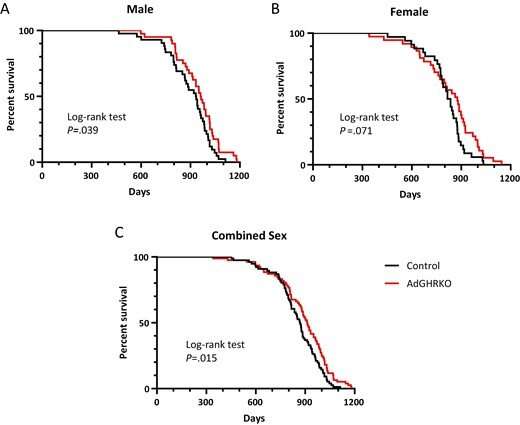
The results, which were recently published in the journal Endocrinology, where it received the distinction of a Featured Article, showed that disrupting the growth hormone receptor (GHR) gene in fat cells improved  at an advanced age and increased lifespan in male mice. The mice also had increased fat mass, reduced circulating levels of insulin, C-peptide, adiponectin, resistin, and improved frailty scores with increased grip strength at advanced ages. The researchers found that approximately 23% lifespan extension in male mice that have no GH action is due to GHR disruption in fat cells. In a previous report, they determined that approximately 19% of the lifespan extension is due to GH action in muscle, and that females benefited less from GHR disruption.

Study finds disruption of the growth hormone receptor gene in adipose tissue results in increased lifespan in mice
Kaplan-Meier survival plots for AdGHRKO mice. AdGHRKO mice (red) and littermate controls (black) are shown for males (A, top left), females (B, top right), and combined sexes (C, bottom). P values are provided for log-rank tests and shown in bold if statistical significance is reached (P = less than .05). AdGHRKO, adipocyte-specific growth hormone receptor knockout. Credit: Endocrinology (2022). DOI: 10.1210/endocr/bqac129

The study was conceived as investigators considered a line of mice developed in Kopchick's lab here at Ohio University that has been used for more than 25 years to study healthy aging. This line of mice, called  receptor knockout (GHRKO) mice, have no GH action in their bodies. They live longer, have enhanced insulin sensitivity and are protected from multiple age-related diseases.

"In fact, this mouse, which is completely GH-insensitive because it lacks the receptor for GH (GHR), holds the record for the longest-lived laboratory mouse, living one week shy of five years versus control mice, which live about two to two and a half years," List said. "We also know that GH acts on  so we wanted to find out what happens if we take away GHR only in these cells while leaving GHR intact in all other cells and tissues."

After removing the GH receptor in adipocytes in mice, the investigators allowed the mice to live normally and analyzed if the alteration impacted their metabolism and lifespan, looking at adiposity, cytokines/adipokines, glucose homeostasis, frailty, and lifespan in aging mice of both sexes.

According to the study, the data demonstrate that removal of GH's action, even in a single tissue, is sufficient for observable health benefits that promote long-term health, reduce frailty, and increase longevity.

"This study is important because it tells us that some of the not-so-beneficial health effects of GH occur in  and that GH is not an anti-aging drug; if anything it promotes aging," List said.

More information: Edward O. List et al, Disruption of Growth Hormone Receptor in Adipocytes Improves Insulin Sensitivity and Lifespan in Mice, Endocrinology (2022). DOI: 10.1210/endocr/bqac129

https://medicalxpress.com/news/2022-10-disruption-growth-hormone-receptor-gene.html

Catholic Bishops Blast Biden for Promoting Abortion: 'Single-Minded Extremism is Gravely Wrong'

 U.S. Catholic bishops issued a message condemning President Joe Biden’s “single-minded extremism” on abortion this week, telling the supposedly Catholic politician that killing unborn babies is “gravely wrong.”

The Catholic News Agency reports the message came from Archbishop William Lori of Baltimore on Tuesday in response to Biden naming a radical pro-abortion bill his top priority for 2023. Another archbishop recently described the legislation as “child sacrifice,” saying it’s what “one would expect from a devout Satanist, not a devout Catholic.”

Lori, chair of the Committee on Pro-Life Activities for the U.S. Conference of Catholic Bishops, said Biden’s abortion advocacy is “tragic” and “gravely wrong” because babies in the womb are valuable human beings, according to the report.

“The president is gravely wrong to continue to seek every possible avenue to facilitate abortion, instead of using his power to increase support and care to mothers in challenging situations,” Lori said. “This single-minded extremism must end, and we implore President Biden to recognize the humanity in preborn children and the genuine life-giving care needed by women in this country.”

The bill that Biden supports would force states to legalize abortions for basically any reason up to birth and force taxpayers to pay for them. Nicknamed the Abortion Without Limits Up to Birth Act, the legislation also would end parental consent for minors and jeopardize conscience protections for doctors and nurses who refuse to abort unborn babies.

Lori told Biden that abortions are violent, tragic and unnecessary and pastors see the devastating effects of abortion daily. He urged the president to work with the Catholic Church to support families in need, including children in the womb.

“… abortion is a violent act which ends the life of preborn children and wounds untold numbers of women,” the archbishop said. “The Catholic Church wishes to continue in our work with our government and leaders to protect the right to life of every human being and to ensure that pregnant and parenting mothers are fully supported in the care of their children before and after birth.”

Biden professes to be a devout Catholic, but he is the most pro-abortion president in U.S. history. His administration recently began allowing dangerous abortion drugs to be sold through the mail without a medical exam, and forcing taxpayers to fund travel for military members to get abortions. Biden also wants to force VA hospitals to abort unborn babies and end the Hyde Amendment.

Last week, he said his top priority for 2023 is a bill to “codify Roe,” a ruling that led to the deaths of more than 63 million unborn babies in abortions.

Some bishops say Biden should not receive communion because he is “not in communion with the Catholic faith.”

“Mr. Biden’s apostasy on the abortion issue is only the most repugnant example. He’s not alone. But in a sane world, his unique public leadership would make — or should make — public consequences unavoidable,” Archbishop Charles Chaput said last week. “When you freely break communion with the Church of Jesus Christ and her teachings, you can’t pretend to be in communion when it’s convenient.”

The Catholic Church teaches that every human being is valuable, from conception to natural death.

https://www.lifenews.com/2022/10/26/catholic-bishops-blast-biden-for-promoting-abortion-his-single-minded-extremism-is-gravely-wrong/

Gene involved in neuronal structure and function may protect against Alzheimer's

 The overexpression of a gene tied to cell division and the structure and function of neurons may prevent and protect against cognitive decline in both mice and humans with Alzheimer's disease (AD), according to a new study by scientists at the University of Colorado Anschutz Medical Campus.

The gene, Kinesin-5 or KIF11, does this despite the presence of amyloid beta (Abeta), the main component of plaques in the brains of those with AD. Scientists have traditionally targeted the plaques when looking for treatments for the fatal disease. In this case, they went around them.

The study was published online last week in the journal iScience.

"Overexpressing KIF11 in mice did not affect the amyloid levels in the brain," said the study's co-senior author Huntington Potter, PhD, professor of neurology and director of the University of Colorado Alzheimer's and Cognition Center and of Alzheimer's research at the Linda Crnic Institute for Down Syndrome at the University of Colorado School of Medicine. "Yet they were still cognitively normal despite the plaques. This is one of the best indications that you can maintain cognition without getting rid of the plaques."

KIF11 is a motor protein best known for its role in mitosis or cell division in non-neuronal cells. But it also plays a critical role in the formation of the dendrites and dendritic spines of neurons, which are used to communicate with other neurons and are important for learning and memory. Yet the main component of Alzheimer's plaques, Abeta, can inhibit KIF11 and cause damage to these structures.

The researchers found that overexpressing the gene in mice with AD led to improved performance on cognitive tests compared to AD mice with normal levels of KIF11. Then they analyzed genetic data from human AD patients provided by the Religious Orders Study and the Rush Memory and Aging Project (ROS/MAP) at Rush University in Chicago. They wanted to know if naturally occurring variations in KIF11 levels correlated with better cognitive performance in adults with or without amyloid plaques.

"Our results from analyzing the human data indicate that higher levels of KIF11 correlate with better cognitive performance in a cohort of older adults with amyloid pathology," said the study's lead author Esteban Lucero, PhD, from the University of Colorado School of Medicine.

"Thus, our results suggest that higher KIF11expression levels may partially prevent cognitive loss during the course of AD in humans, which aligns with our findings regarding the role of KIF11 in animal models of AD," Lucero said.

Potter and co-senior author Heidi Chial, PhD, assistant professor of neurology and director of grant strategy and development at the University of Colorado Alzheimer's and Cognition Center, said this information paves the way for researchers to begin testing new or existing drugs that can safely create this effect in humans.

"Many current experimental treatments for AD have focused on reducing Abeta production or on increasing the clearance of Abeta plaques," Chial said. "Most of these approaches have failed to prevent or reverse cognitive decline in clinical trials. Clearly, alternative approaches to the development of AD therapeutics are needed."


Story Source:

Materials provided by University of Colorado Anschutz Medical Campus. Original written by David Kelly. Note: Content may be edited for style and length.


Journal Reference:

  1. Esteban M. Lucero, Ronald K. Freund, Alexandra Smith, Noah R. Johnson, Breanna Dooling, Emily Sullivan, Olga Prikhodko, Md. Mahiuddin Ahmed, David A. Bennett, Timothy J. Hohman, Mark L. Dell’Acqua, Heidi J. Chial, Huntington Potter. Increased KIF11/kinesin-5 expression offsets Alzheimer Aβ-mediated toxicity and cognitive dysfunctioniScience, 2022; 25 (11): 105288 DOI: 10.1016/j.isci.2022.105288

Potential to block nerve loss in neurodegenerative diseases

 Two new studies from Washington University School of Medicine in St. Louis support development of a broadly applicable treatment for neurodegenerative diseases that targets a molecule that serves as the central executioner in the death of axons, the wiring of the nervous system.

Blocking this molecular executioner prevents axon loss, which has been implicated in many neurodegenerative diseases, from peripheral neuropathies to Parkinson's disease, and glaucoma to amyotrophic lateral sclerosis (ALS).

The new studies, both published Oct. 26 in the Journal of Clinical Investigation, reveal surprising details about how the molecule -- called SARM1 -- triggers axon death that underlies the development of neurodegenerative diseases. The research also points to new therapeutic approaches for diseases defined by axon loss.

"We desperately need treatments for neurodegenerative diseases," said co-senior author Jeffrey Milbrandt, MD, PhD, the James S. McDonnell Professor and head of the Department of Genetics. "With the evidence of SARM1's central role in these diseases, we're very interested in finding ways to block this molecule -- whether with small molecule inhibitors or gene therapy techniques. Our latest research suggests we also may be able to interfere with its ability to drive damaging neuroinflammation. We're hopeful this work will lead to effective new therapies across a range of neurodegenerative and neuroinflammatory diseases."

In 2017, Milbrandt and co-senior author Aaron DiAntonio, MD, PhD, the Alan A. and Edith L. Wolff Professor of Developmental Biology, discovered that SARM1 is an enzyme that can promote neurodegeneration. Soon after, they co-founded a startup company called Disarm Therapeutics to boost the development of drug compounds that inhibit SARM1 for the treatment of diseases characterized by axon degeneration. In 2020, Disarm Therapeutics was acquired by Eli Lilly and Company to further the development of SARM1-targeted therapies for neurodegenerative diseases.

In healthy neurons, SARM1 is always switched off. But after injury or due to disease, SARM1 becomes active. Activated SARM1 is an arsonist -- burning so much cellular energy that the axons can't survive. This energy crisis triggers axons to disintegrate.

To understand more about SARM1's role in triggering axon destruction, the researchers studied a mysterious and extremely rare progressive neuropathy syndrome -- so rare, it lacks a name. This rare disease turned out to be a good model for understanding the role of the immune system in neuroinflammatory conditions generally. Sequencing patient genomes, the researchers found that the axon loss was caused by genetic errors in the gene NMNAT2, whose normal function keeps SARM1 turned off. Due to these genetic errors, SARM1 is constantly activated, which triggers axon destruction. The researchers used the CRISPR gene-editing technique to reproduce these mutations in mice. Like people with the syndrome, these mice survived to adulthood but had worsening motor dysfunction, loss of peripheral axons and, importantly, an infiltration of immune cells called macrophages.

The researchers were surprised to find that reducing the number of macrophages reversed the axon loss and disease symptoms in the mice. The study suggests that SARM1 not only contributes directly to axon loss but also plays a role in driving neuroinflammation that only serves to compound the problems. The findings also suggest that some neurodegenerative conditions could be treated with immune modulating drugs that block macrophages or other inflammatory immune cells.

In the second paper, the researchers investigated the possible role of SARM1 in Charcot-Marie-Tooth disease type 2a, a common form of inherited peripheral neuropathy and a good model to study axon loss generally. Patients with this disease have progressive loss of motor and sensory axons and develop difficulty walking, muscle weakness, and tingling or burning sensations in the hands and feet. The disease is caused by a mutation in an important protein in mitochondria, the energy factories of cells. The mutation, in a protein called mitofusin2, impairs the normal function of mitochondria. Much research has focused on the abnormal mitochondria, assuming they must be the root of the problem in this disease.

Surprisingly, the researchers found that deleting SARM1 in a rodent model of Charcot-Marie-Tooth disease type 2a stopped most of the problems the animals exhibited -- independent of the diseased mitochondria. Eliminating SARM1 blocked or slowed axon death, muscle atrophy, mitochondrial abnormalities and problems with neuromuscular junctions, where the neurons interface with muscle. Even with the mutant mitofusin2 protein present, deleting SARM1 protected the mitochondria from further degradation and dysfunction.

"When we block SARM1, we not only protect the axons, we get much healthier mitochondria," DiAntonio said. "This was a complete surprise, but we are hopeful it could be relevant in many neurodegenerative diseases where mitochondrial damage is central, such as Parkinson's disease, as many neurodegenerative diseases have a component of mitochondrial dysfunction."

Milbrandt and DiAntonio are co-founders, scientific advisory board members and shareholders of Disarm Therapeutics, a wholly owned subsidiary of Eli Lilly and Company. Disarm Therapeutics and Eli Lilly are developing SARM1-targeted therapies for neurodegenerative diseases.


Story Source:

Materials provided by Washington University School of Medicine. Original written by Julia Evangelou Strait. Note: Content may be edited for style and length.


Journal References:

  1. Yurie Yamada, Amy Strickland, Yo Sasaki, A. Joseph Bloom, Aaron DiAntonio, Jeffrey Milbrandt. A SARM1/mitochondrial feedback loop drives neuropathogenesis in a Charcot-Marie-Tooth disease type 2A rat modelJournal of Clinical Investigation, 2022; DOI: 10.1172/JCI161566
  2. Caitlin B. Dingwall, Amy Strickland, Sabrina W. Yum, Aldrin Kay-Yuen Yim, Jian Zhu, Peter L. Wang, Yurie Yamada, Robert E. Schmidt, Yo Sasaki, A. Joseph Bloom, Aaron DiAntonio, Jeffrey Milbrandt. Macrophage depletion blocks congenital SARM1-dependent neuropathyJournal of Clinical Investigation, 2022; DOI: 10.1172/JCI159800

Engineers light the way to nerve-operated prosthetics of the future

 Biomedical and electrical engineers at UNSW Sydney have developed a new way to measure neural activity using light -- rather than electricity -- which could lead to a complete reimagining of medical technologies like nerve-operated prosthetics and brain-machine interfaces.

Professor François Ladouceur, with UNSW's School of Electrical Engineering and Telecommunications, says the multi-disciplinary team has just demonstrated in the lab what it proved theoretically shortly before the pandemic: that sensors built using liquid crystal and integrated optics technologies -- dubbed 'optrodes' -- can register nerve impulses in a living animal body.

Not only do these optrodes perform just as well as conventional electrodes -- that use electricity to detect a nerve impulse -- but they also address "very thorny issues that competing technologies cannot address," says Prof. Ladouceur.

"Firstly, it's very difficult to shrink the size of the interface using conventional electrodes so that thousands of them can connect to thousands of nerves within a very small area.

"One of the problems as you shrink thousands of electrodes and put them ever closer together to connect to the biological tissues is that their individual resistance increases, which degrades the signal-to-noise ratio so we have a problem reading the signal. We call this 'impedance mismatch'.

"Another problem is what we call 'crosstalk' -- when you shrink these electrodes and bring them closer together, they start to talk to, or affect each other because of their proximity."

But because optrodes use light and not electricity to detect neural signals, the problems of impedance mismatch is redundant and crosstalk minimised.

"The real advantage of our approach is that we can make this connection very dense in the optical domain and we don't pay the price that you have to pay in the electrical domain," Prof. Ladouceur says.

In vivo demonstration

In research published recently in the Journal of Neural Engineering, Prof. Ladouceur and fellow researchers at UNSW wanted to show that they could use optrodes to accurately measure the neural impulses as they travel along a nerve fibre in a living animal.

Scientia Professor Nigel Lovell, who heads the Graduate School of Biomedical Engineering and is Director of the Tyree Foundation Institute of Health Engineering, was part of the research team that sought to demonstrate this in the lab.

He says the team connected an optrode to the sciatic nerve of an anaesthetised animal. The nerve was then stimulated with a small current and the neural signals were recorded with the optrode. Then they did the same using a conventional electrode and a bioamplifier.

"We demonstrated that the nerve responses were essentially the same," says Prof. Lovell. "There's still more noise in the optical one, but that's not surprising given this is brand new technology, and we can work on that. But ultimately, we could identify the same characteristics by measuring electrically or optically."

New dawn for prosthetics

So far the team has been able to show that nerve impulses -- which are relatively weak and measured in microvolts -- can be registered by optrode technology. The next step will be to scale up the number of optrodes to be able to handle complex networks of nervous and excitable tissue.

Prof. Ladouceur says at the beginning of the project, his colleagues asked themselves, how many neural connections does a man or woman need to operate a hand with a degree of finesse?

"That you can pick up an object, that you can judge the friction, you can apply just the right pressure to hold it, you can move from A to B with precision, you can go fast and slow -- all these things that we don't even think about when we perform these actions. The answer is not so obvious, we had to search quite a bit in the literature, but we believe it's about 5000 to 10,000 connections."

In other words, between your brain and your hand there is a bundle of nerves that travels down from your cortex and eventually divides into those 5000 to 10,000 nerves that control the delicate operations of your hand.

If a chip with thousands of optical connections could connect to your brain, or some place in the arm before the nerve bundle separates, a prosthetic hand could potentially be able to function with much the same ability as a biological one.

That's the dream, anyway, and Prof. Ladouceur says there are likely decades of further research before it's a reality. This would include developing the ability for optrodes to be bidirectional. Not only would they receive and interpret signals from the brain on the way to the body, they could receive feedback in the form of neural impulses going back to the brain.

The long game: brain-machine interface

Neural prosthetics isn't the only space that optrode technology has the potential to redefine. Humans have long fantasised about integrating technology and machinery into the human body to either repair or enhance it.

Some of this is now a reality, such as Cochlear implants, pacemakers and cardiac defibrillators, not to mention smart watches and other tracking devices giving continual biofeedback.

But one of the more ambitious goals in biomedical engineering and neuroscience is the brain-machine interface that aims to connect the brain to not only the rest of the body, but potentially the world.

"The area of neural interfacing is an incredibly exciting field and will be the subject of intense research and development over the next decade," says Prof. Lovell.

While this is more fiction than fact right now, there are many biotech companies taking this very seriously. Entrepreneur Elon Musk was one of the co-founders of Neuralink that aims to create brain-computer interfaces with the potential to help people with paralysis as well as incorporating artificial intelligence into our brain activities.

The Neuralink approach uses conventional wire electrodes in its devices so it must overcome impedance mismatch and crosstalk -- among many other challenges -- if they are to develop devices that host thousands, if not millions, of connections between the brain and the implanted device. Recently Mr Musk was reported as being frustrated at the slow pace in developing the technology.

Prof. Ladouceur says time will tell whether Neuralink and its competitors succeed in removing these obstacles. However, given that implantable, in vivo devices that capture neural activity are currently constrained to about 100 or so electrodes, there is still a long way to go.

"I'm not saying that it's impossible, but it becomes really problematic if you were to stick to standard electrodes," Prof. Ladouceur says.

"We don't have these problems in the optical domain. In our devices, if there is neural activity, its presence influences the orientation of the liquid crystal which we can detect and quantify by shining light on it. It means we don't extract current from the biological tissues as the wire electrodes do. And so the biosensing can be done much more efficiently."

Now that the researchers have shown that the optrode method works in vivo, they will shortly publish research that shows the optrode technology is bidirectional -- that it can not only read neural signals, but can write them too.


Story Source:

Materials provided by University of New South Wales. Original written by Lachlan Gilbert. Note: Content may be edited for style and length.


Journal Reference:

  1. Amr Al Abed, Yuan Wei, Reem M Almasri, Xinyue Lei, Han Wang, Josiah Firth, Yingge Chen, Nathalie Gouailhardou, Leonardo Silvestri, Torsten Lehmann, François Ladouceur, Nigel H Lovell. Liquid crystal electro-optical transducers for electrophysiology sensing applicationsJournal of Neural Engineering, 2022; 19 (5): 056031 DOI: 10.1088/1741-2552/ac8ed6