Search This Blog

Friday, February 17, 2023

The AMA Said 'Trust Your Doctor' On Smoking

 by Kevin Homer via The Brownstone Institute,

The American Medical Association (AMA) urges physicians to promote COVID-19 vaccines and bivalent boosters

The AMA even supplies members with social media talking points and strategies to deal with vaccine detractors

It is not the first time that my profession has endorsed a product that may be hazardous to your health.

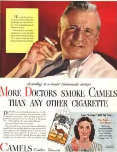
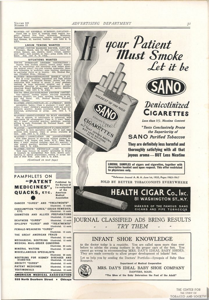
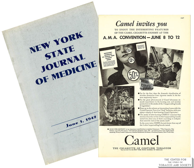
For most of the 20th century, the AMA turned a blind eye toward the dangers of tobacco use. During the 1930s, 40s and 50s, tobacco companies paid handsomely to advertise cigarettes in AMA’s journal, JAMA. In a 1948 editorial minimizing the ill effects of smoking and justifying tobacco advertising in its publications, JAMA noted that “cigarette business is a tremendous business,” as if the size of the bottom line can mitigate a conflict for an organization founded for the “betterment of public health.”

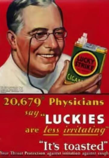
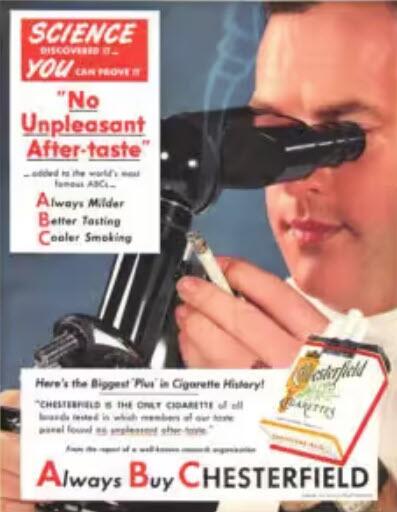
The connection between smoking and lung cancer was recognized early in the century. At the same time, the AMA became increasingly dependent on money generated by tobacco sales. Tobacco companies sponsored meetings of medical societies, setting up their booths alongside exhibitions of the latest medical treatments. Free cartons were distributed at physician meetings. Cigarette makers even paid for publication of pseudoscientific reports claiming the health benefits of their products.  

Doctors who opposed smoking faced ridicule from their colleagues. Dr. Alton Ochsner, a renowned surgeon and sentinel voice warning of the dangers of tobacco, began publishing on the connection between smoking and lung cancer in the early 1940s. His 1954 book Smoking and Cancer: A Doctor’s Report was negatively reviewed in prominent medical journals, characterized as a medieval model of logic that belongs in the nonscience section of a library. Prior to his appearance on Meet the Press, Dr. Ochsner was told he could not discuss the relationship between smoking and lung cancer on air.

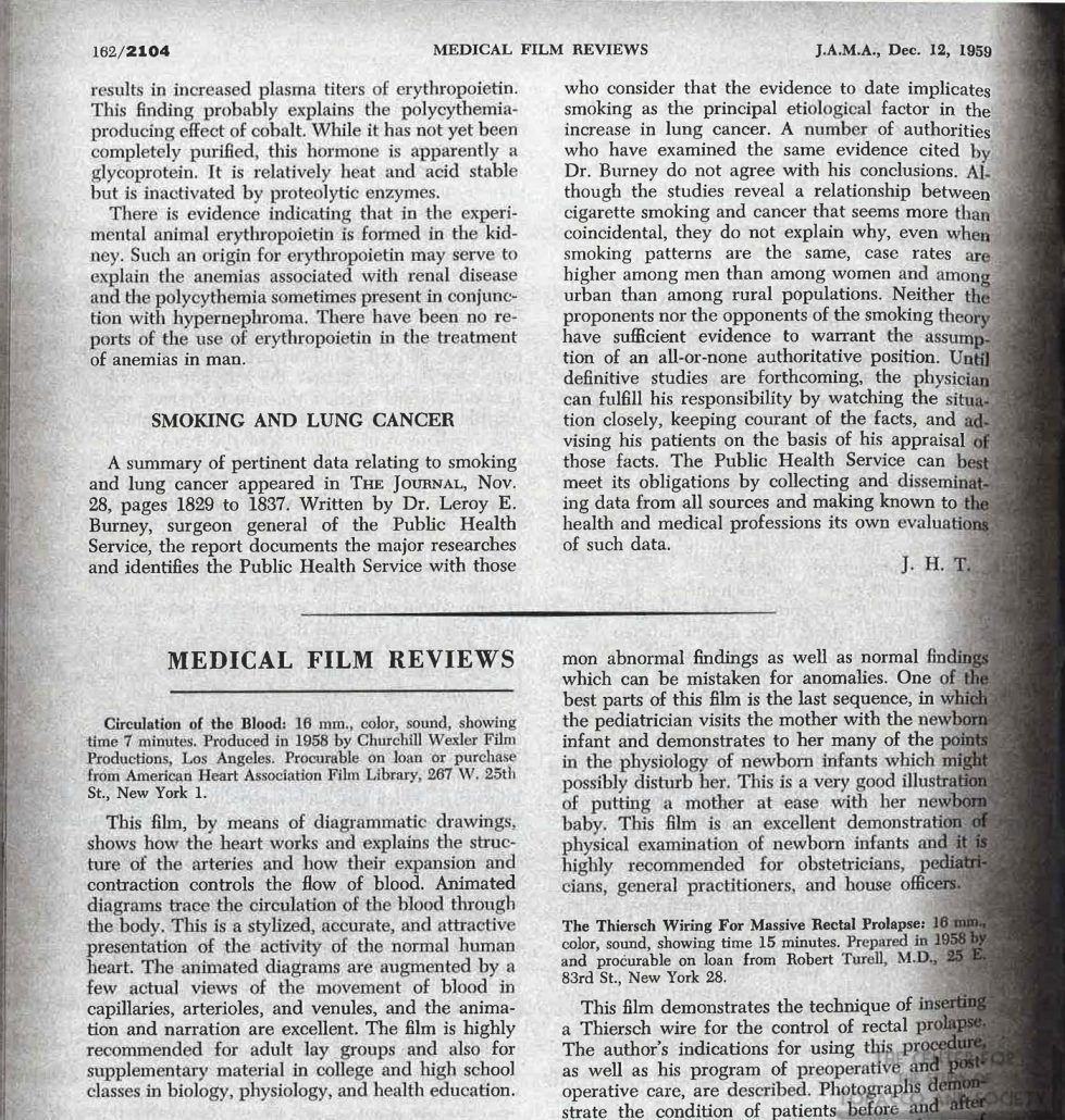
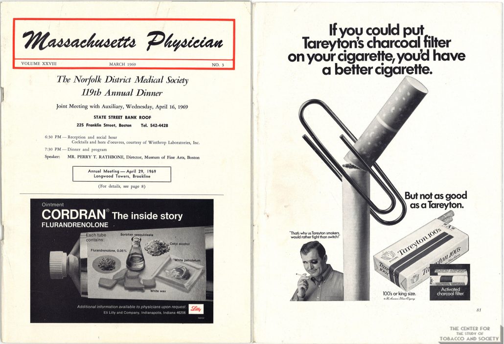
Yet the mounting evidence was hard to ignore. In 1954, JAMA stopped accepting cigarette advertisements and published an editorial rebuking tobacco company advertising practices. But five years later, a JAMA editorial was still skeptical of the evidence linking smoking to cancer, and a 1961 Nebraska State Medical Journal editorial dismissed the evidence as merely “statistical.” Tobacco companies continued to sponsor state medical meetings as late as 1969. By then most people were aware of the dangers of smoking.

In 1964, the Surgeon General concluded that cigarette smoking causes lung cancer and other life-limiting health conditions. The next year, a warning label was required on packages of cigarettes. By 1971, the government banned cigarette advertisements on television and radio. Instead of taking the lead against an obvious threat to public health, the AMA asked for time and money to study the effects of tobacco. 

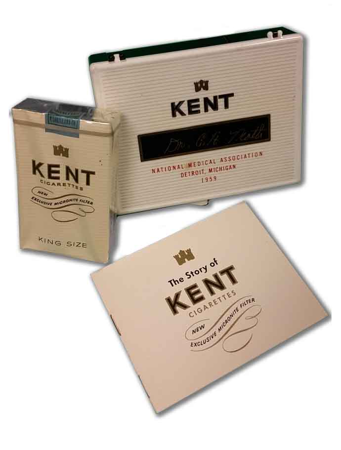
Between 1964 and 1976, the AMA received more than $20 million from the tobacco industry to fund research. Instead of using the money for smoking cessation programs, many of the funded studies focused on ways to make a safer cigarette. To keep money flowing into its Education and Research Foundation the AMA delayed, stating in a confidential 1971 report that, “AMA is not prepared to make any statement regarding termination of the smoking-health research program.” The report went on to complain that tobacco companies are “in arrears on 1970 contributions.” The dependency on tobacco money created a political alliance between doctors and cigarette makers as their lobbyists joined forces in Washington. 

The delay benefitted tobacco sales and maintained the AMA’s “research” payments, but it angered Dr. Ochsner, who accused the AMA of being derelict. The AMA called Dr. Ochsner’s position “extreme.” But name-calling could not stall the inevitable conclusion any longer. In 1978 the AMA finally agreed with what most people had already realized: smoking causes lung cancer, and many other health problems. The romance with big tobacco was over.

Or was it?

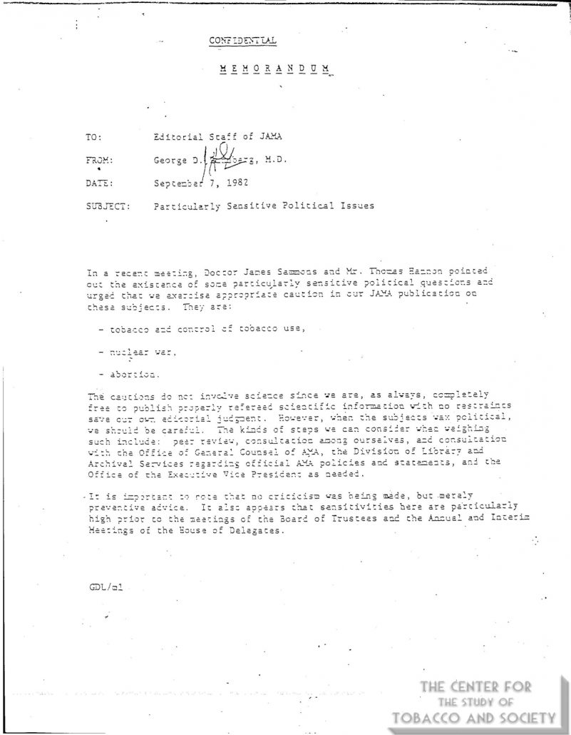
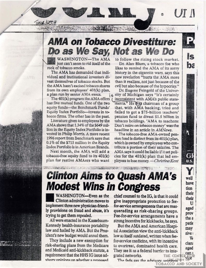
As late as 1982, JAMA publications were warned to steer clear of “politically sensitive” topics like tobacco use. After most of a century of being on the tobacco dole, the AMA could not make a clean break. The AMA portfolio contained investments in tobacco companies until the late 1990s. 

In 1998, the tobacco industry settled lawsuits filed by state governments with a massive Master Settlement Agreement. In exchange for perpetual annual payments and tight regulatory control, the tobacco industry could continue to sell its products protected from future lawsuits brought by participating states and jurisdictions.  

But who really benefitted from the Tobacco Settlement? Only 2.6 percent of the money has been used for smoking prevention and cessation programs. Some states have used the tobacco money to fill budget gaps. South Carolina gave money to tobacco farmers affected by a drop in prices. Altria Group, a global tobacco company, is on the US News & World Report 10 best-performing stocks list. Altria, Phillip Morris, and British American Tobacco have all grown annual dividends consecutively since the settlement. According to Dr. Ed Anselm, “The most addictive thing about tobacco is money.”

Tobacco use remains the number one preventable cause of death in the United States. In the first fifty years after the Surgeon General’s 1964 report, more than 20 million Americans died of smoking. How many of these deaths would have been prevented if doctors had not been conflicted by financial entanglements with the tobacco industry?

Money blinds objectivity. When money drives decisionscontroverting evidence is ignoreddissenting voices are ridiculedopen debate is suppressedtalking points are distributedconclusions are delayed, and people die from a product with liability protection

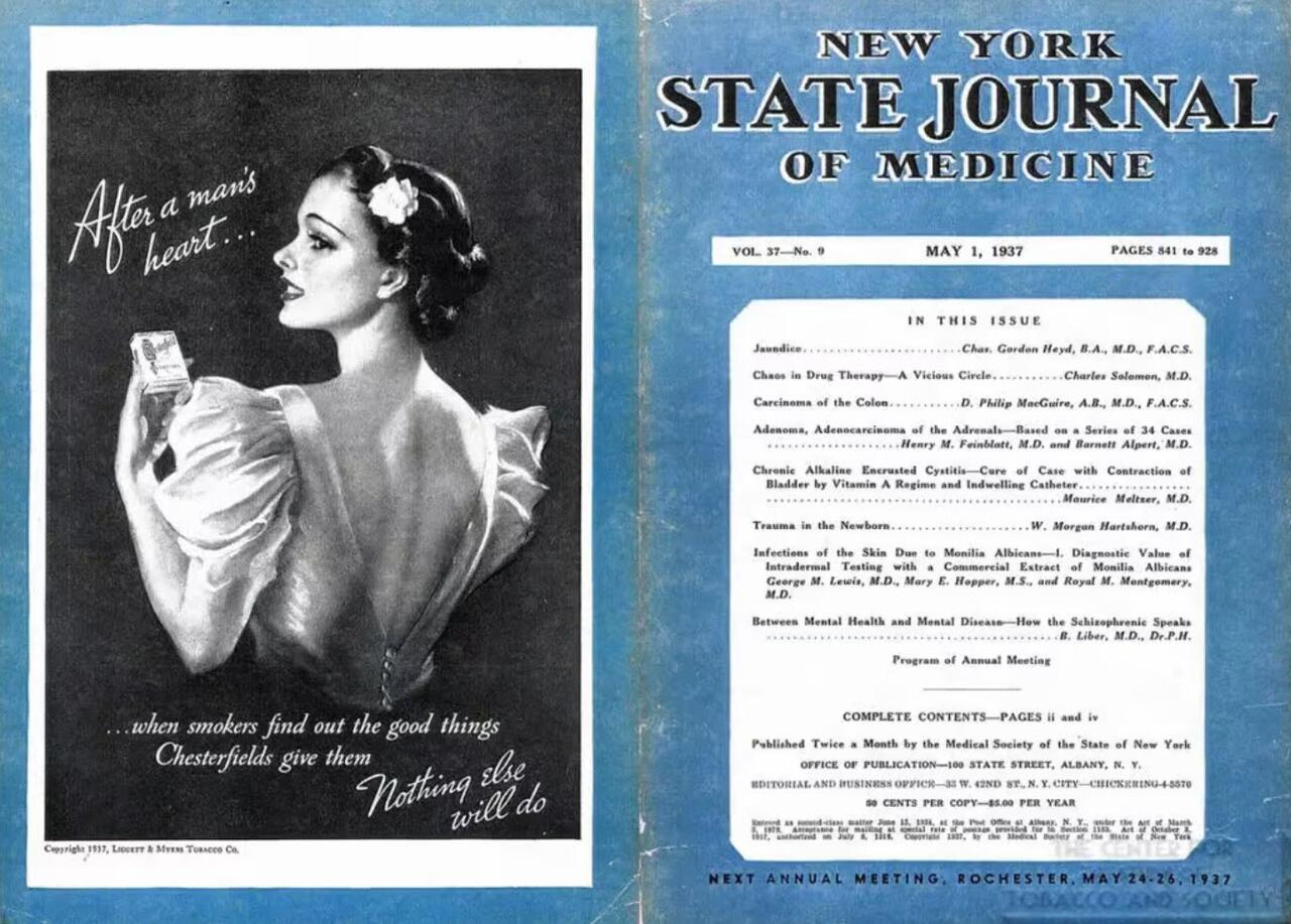
The New York State Journal of Medicine published a retrospective of tobacco’s relationship to medicine in its December 1983 issue. Flipping through the pages is enlightening.  Surrounding the articles describing the greed and politics of Big Tobacco are advertisements from medicine’s new love—Big Pharma.  Doctors have exchanged one bedfellow for another.

By endorsing irrelevant COVID-19 vaccines and poorly tested bivalent boosters, the AMA is pushing a product without concern for its potential negative health effects. Like before, the medical profession lags behind public opinion. According to recent Rasmussen Reports, 7 percent of vaccinated individuals report a major side effect, and nearly half of Americans believe that COVID-19 vaccines have caused unexplained deaths, about the same proportion who believed that smoking caused cancer in the 1960s while the AMA was studying the issue.

conflicted profession cannot honestly evaluate data. Nowadays, the pharmaceutical business is a tremendous business. An organization benefitting from product sales cannot be trusted to evaluate that product. 

If doctors could not recognize the health dangers of tobacco for most of the last century, why should we trust them when they say novel vaccines are safe and effective?

https://www.zerohedge.com/medical/ama-said-trust-your-doctor-smoking

20 Women Sexually Abused At Epstein Properties Paid Through JPMorgan Accounts: Filing

 by Zachary Stieber via The Epoch Times (emphasis ours),

At least 20 women who were victims of sex trafficking and sexual abuse at Jeffrey Epstein properties were paid through JPMorgan Chase accounts, according to a new court filing.

The women were allegedly abused and trafficked at properties in the U.S. Virgin Islands, New York, and elsewhere between 2003 and 2019.

The women received payments that totaled, collectively, more than $1 million, according to the U.S. Virgin Islands Department of Justice, which entered the filing on Feb. 15.

Epstein also withdrew more than $775,000 in cash over that time frame from JP Morgan accounts, especially significant as Epstein was known to pay for ‘massages,’ or sexual encounters, in cash,” the filing states. “Financial information also reflects payments drawn from JP Morgan accounts of nearly $1.5 million to known recruiters, including to the MC2 modeling agency, and another $150,000 to a private investigative firm.”

Epstein was facing sex trafficking charges when he died in jail in 2019. New York City’s medical examiner ruled the death a suicide. Epstein had pleaded guilty to soliciting a minor for prostitution in Florida in 2008.

Then-U.S. Virgin Islands Attorney General Denise George brought a case against JPMorgan Chase in late 2022, alleging the company helped Epstein carry out crimes including sex trafficking. George was fired shortly after, but the case is moving forward.

The new filing is a less redacted version of the first amended complaint, which had been entered into the docket previously.

‘Extremely High-Risk Client’

Evidence shows JPMorgan officials were aware Epstein was an “extremely high-risk client” but decided to keep servicing his accounts “because of his vast wealth and connections with other high net worth individuals,” according to the complaint.

The company’s global corporate security division, for instance, reviewed media articles that detailed the charges against Epstein in Florida. JPMorgan decided to keep doing business with Epstein, though it did label him as “high risk.”

In a 2010 internal email, the division flagged “new allegations of an investigation related to child trafficking” and wondered, “are you still comfortable with this client who is now a registered sex offender.”

In January 2011, the company’s compliance director asked for reapproval of the relationship due to the new allegations of human trafficking. “I thought we did that in approving a $50 million new line of credit last month?” another employee responded.

A review completed that month led to the conclusion that no “material updates” had been identified. It also noted that Jes Staley, a top JPMorgan official, had spoken with Epstein about the alleged human trafficking. Epstein said there was no truth to the allegations or evidence supporting them.

Just two months later, the security division reported how Epstein had settled a dozen lawsuits with alleged victims and that Epstein was connected to Jean Luc Brunel, owner of the MC2 Model Management agency. The agency received $1 million from Epstein in 2005, according to the vision, which stated, “It is unknown if the money was given as a secret investment or payment for services as a procurer.”

https://www.zerohedge.com/markets/20-women-sexually-abused-jeffrey-epstein-properties-were-paid-through-jpmorgan-accounts

Daily rhythm detected for cerebral blood flow in stroke patients

 Strokes are a leading cause of morbidity and mortality in the United States, as well as around the world. Various environmental and biological factors are known to affect the risks and outcomes of strokes. A new study led by investigators from Brigham and Women's Hospital, a founding member of the Mass General Brigham healthcare system, analyzed the cerebral blood flow (CBF) regulation of individuals who had experienced strokes.

The team found that cerebral autoregulation (CA)—one of the key processes to maintain sufficient blood supply to the brain—displayed a daily  in , with more degraded regulation during nighttime and morning hours, as compared to that in the afternoon hours. Their results, which are published in the Journal of Cerebral Blood Flow and Metabolism, are relevant for  planning during  recovery.

"The care and course of actions done after a stroke are essential for optimal rehabilitation. Our study suggests that the daily rhythm of CBF regulation in stroke patients may be highly relevant to managing an individual's activity and stroke recovery," said senior author, Kun Hu, Ph.D., of the Medical Biodynamics Program in the Brigham's Division of Sleep and Circadian Disorders.

"Exercise and surgery post-stroke could be more optimal when scheduled during afternoon hours, as this is when dynamic CA functions more effectively. These results may improve our understanding of a vulnerable time window for the cerebrovascular system and help guide daily activity and personal care during stroke recovery, which could improve health outcomes for patients who have had a stroke."

Stable CBF is a necessary component for normal brain function. Dramatic changes in CBF can cause increased cranial pressure and brain tissue damage. Thus, a process like CA, which helps maintain relatively stable CBF through constriction and dilation of blood vessels in the brain, particularly during changes in an individual's blood pressure (BP), is crucial.

Currently, there is a gap in knowledge concerning the daily variance of CA in stroke patients. Generally, the daily rhythms of physiological functions can be controlled by external behaviors like food intake, sleep, and exercise, as well as the internal circadian clock. This study is among the first to examine the potential variation of CA in the stroke population.

The research team observationally studied 28 participants being treated in a hospital in São Paulo, Brazil after experiencing a stroke. They received thrombolysis within 5 hours of the onset of their symptoms. After undergoing this procedure, the participants' CA was assessed over the course of 48 hours at various time points by examining the relationship between temporal changes in blood pressure and cerebral blood flow velocity of the middle cerebral artery.

Analysis of the results showed evidence of differing cerebral  flow regulation during various times of the day, especially when  and pressure fluctuated at large time scales or low frequencies < 0.05 Hz. In particular, a more degraded regulation motif was seen during the nighttime and  when compared to the afternoon. This dysregulation interval coincides with the increased prevalence of recurrent and first-ever stroke events during morning times.

While these results are promising, the researchers identified future avenues to assist in creating a greater understanding of the regulation mechanism.

"Interestingly, the daily rhythm of CA was present in both stroke and non-stroke sides of the brain, suggesting the factor(s) driving the rhythm should affect CBF regulation globally," said first author Daniel Abadjiev, of the Medical Biodynamics Program in the Brigham's Division of Sleep and Circadian Disorders.

"To better understand underlying mechanisms, future studies should consider more frequent assessments across the 24-hour cycle, an increase in patient sample size, inclusion of non-stroke controls, and monitoring of the daily activity rhythms like sleep and exercise as well as intrinsic circadian rhythm among the participants."

"This study demonstrates that a daily rhythm does exist for stroke patients," said Hu. "Rehab plans should look to identify the daily rhythm and design a strategy that makes use of optimal CA. The long-term goal is to see if we can further control the rhythm of CA by manipulating an individual's daily behavioral cycles or endogenous circadian clock in order to deliver more personalized medicine and improve their recovery."

More information: Daniel S Abadjiev et al, Daily rhythm of dynamic cerebral autoregulation in patients after stroke, Journal of Cerebral Blood Flow & Metabolism (2023). DOI: 10.1177/0271678X231153750


https://medicalxpress.com/news/2023-02-daily-rhythm-cerebral-blood-patients.html

Pharmacists based in care homes found to be safer for residents

 Basing specialist pharmacists in care homes can help make residents safer by cutting potential harm from medicines, according to research carried out by University of Leeds academics.

The Care Home Independent Pharmacist Prescriber Study (CHIPPS), which involved researchers in Leeds and across the country, trialed onsite pharmacists in dozens of .

Evidence from the study shows that onsite pharmacists reduce the risk of potential harm to care home residents from medicines, cut down on unnecessary medicines, and ease the burden for GPs.

David Alldred, Professor of Medicines Use and Safety in the the University's School of Healthcare, led the project in Yorkshire.

He said, "Older people living in care homes have complex health needs and are usually prescribed multiple medicines. Our large-scale, high-quality study has shown that specially qualified prescribing pharmacists can safely prescribe for care home residents to ensure they are on the right combination of medicines."

"Importantly, the pharmacists also stopped medicines that were no longer needed or appropriate which reduces medicines burden for residents and is likely to prevent future side-effects. Key to success was the pharmacists building relationships and working in partnership with GPs, care home staff and residents."

According to the study, which was published in the BMJ, care home residents are routinely prescribed an average of eight or more medicines.

Medicine use is considered one of the main areas of risk in care homes by both the government and regulatory organizations.

One study cited in the BMJ paper says that 70% of care home residents experienced drug errors daily.

The University of Leicester-led study says that there have been regular calls for interventions to help to improve the management of medicine in care homes.

CHIPPS conducted a randomized controlled trial including 49 care homes across England, Scotland and Northern Ireland.

The study involved 25 care home trained pharmacist independent prescribers who were integrated into care homes to improve medicines management and safety.

It led to researchers determining that embedding pharmacist independent prescribers in care homes is safe, well received by all stakeholders and reduces potential future harm from medicines.

According to the study, independent monitoring of pharmacist activities and review of unexpected  deaths and hospitalizations found no safety concerns.

Cutting unnecessary medicines

Interviews with general practitioners, care home managers, caregivers and residents showed they were highly satisfied with the service provided by the pharmacist independent prescribers.

Analysis of the primary outcome of falls did not show a significant reduction, but potential future harm from medicines was significantly reduced.

University of Aberdeen, Queen's University Belfast, University of East Anglia, NHS Norfolk Waveney and Norwich Clinical Trials also contributed to the research.

Project leader, Professor of Health Services Research and Head of School of Healthcare at Leicester, David Wright said, "I am delighted that the culmination of this six-year program of work, undertaken by an incredible group of researchers, clinicians and patient representatives has produced this result."

"The CHIPPS model of care was very well received by all stakeholders. We believe that this was because both our training and model of delivery focused on the integration of the pharmacist independent prescribers into the  and care home teams."

"This approach was supported by our process evaluation, which found that the intervention appeared most effective when the pharmacists were embedded within their local general practice."

"Furthermore, our pharmacist-led intervention was reported by some GPs to reduce their workload as they no longer had to routinely review and authorize large numbers of repeat medicines and the pharmacist prescribers assumed some of their care home responsibilities."

"The results support expansion of the current pharmacist role in care homes, to include prescribing and frequent visits, as it reduces future harm from medicines and helps care homes to improve their management of medicines."

Professor Richard Holland who co-led the project, added, "Overall, this was a high-quality study which, through independent review of pharmacist care plans, hospitalizations and deaths, did not identify harm resulting from the pharmacist independent prescriber activities."

"This addressed concerns raised by some medical colleagues and a Scottish Ethics Committee at the start of the , when we presented this model of care to them."

"Thus, this study provides important evidence for the development of models of care in care homes which we consider should include  independent prescribers."

More information: Richard Holland et al, Evaluation of effectiveness and safety of pharmacist independent prescribers in care homes: cluster randomised controlled trial, BMJ (2023). DOI: 10.1136/bmj-2022-071883


https://medicalxpress.com/news/2023-02-pharmacists-based-homes-safer-residents.html

Better understanding of how the blood-brain barrier works

 Up to now, the use of models to research the barrier that separates the circulatory from the nervous system has proven to be either limited or extremely complicated. Researchers at ETH Zurich have developed a more realistic model that can also be used to better explore new treatments for brain tumors.

Mario Modena is a postdoc working in the Bio Engineering Laboratory at ETH Zurich. If he were to explain his research on the —the wall that protects our central nervous system from harmful substances in the —to an 11-year-old, he would say, "This wall is important, because it stops the bad guys from getting into the brain." If the brain is damaged or sick, he says, holes can appear in the wall. Sometimes, such holes can actually be useful, for example, for supplying the brain with urgently needed medicine. "So what we are trying to understand is how to maintain this wall, break through it and repair it again."

This wall is also important from a medical perspective, because many diseases of the central  are linked to an injury to the blood-brain barrier. To discover how this barrier works, scientists often conduct experiments on live animals. In addition to such experiments being relatively expensive,  may provide only part of the picture of what is going on in a . Moreover, there are some critics, who question the basic validity of animal testing. An alternative is to base experiments on  that have been cultivated in the laboratory.

Cell–cell communication largely overlooked

The problem with many in-vitro models is that they recreate the blood-brain barrier in a relatively simplified way using blood-vessel-wall cells (endothelial cells). This approach fails to represent the complex structure of the human system and disregards, for instance, the communication between the various cell types. Furthermore, many of these models are static. In other words, the cells are floating in a suspension that is not moving, which implies that  or the  the cells are exposed to in the body are not considered.

There are also dynamic in-vitro models that simulate flow conditions in the body, but the catch here is that the pumps they require make the experimental setup rather complicated. Alongside all these challenges, there is the problem of measurement: it is all but impossible to take high-resolution images of structural changes to the blood-brain barrier in real time while also measuring the barrier's electrical resistance, both of which reflect barrier compactness and tightness.

Achieving a better understanding of how the blood-brain barrier works
The illustration at the top left shows an in-vivo model of the blood-brain barrier, the 

Killing several birds with one stone

If each of these challenges were a bird, Modena's platform would be the proverbial stone that kills them all. Working under Andreas Hierlemann, Modena and his colleagues spent three and a half years developing the open-microfluidic 3D blood-brain barrier model.

To recreate the barrier, the research team took those cell types that naturally make up the blood-brain barrier—microvascular , human astrocytes and human pericytes—and combined them within a single platform. "This strategy allowed us to almost fully replicate the 3D cell structure found in the human body," Modena says. "But what's really exceptional is that we can measure the barrier's permeability while simultaneously mapping morphological changes to the barrier by means of high-resolution time-lapse microscopy."

To facilitate this double act, the researchers deposited entirely  on glass coverslips on both sides of the barrier to measure its permeability, which is reflected in the electrical resistance across the cell barrier. Transparent electrodes offer a decisive advantage over other types of electrodes, which include metal films or wire structures that may interfere with optical detection and high-resolution microscopy.

'Without increasing the complexity'

To mimic the way fluid flows in the body, the researchers realized the microfluidic platform with fluid reservoirs at both ends on a kind of seesaw. Gravity then triggered the flow, which—in turn—generated shear force on the cells. Hierlemann explains the benefit of this setup: "Since we are not using any pumps, we can experiment with multiple model systems simultaneously, for instance in an incubator, without increasing the setup complexity."

In a study, published recently in the journal Advanced Science, the researchers presented and tested their new in vitro blood-brain barrier model. They subjected the barrier to oxygen-glucose deprivation, as happens when someone is having a stroke. "These experiments allowed us to trigger rapid changes in the barrier and demonstrate the platform's potential," Modena says.

Through this study, Modena and his colleagues were able to do more than showing that their new platform is suitable for taking measurements. They also discovered that the barrier's electrical resistance decreases even before it undergoes morphological changes that make it more permeable. "This finding could prove relevant for future research," Modena says.

The team also observed that in control experiments using a static in-vitro model, the barrier was more permeable than in the new dynamic setup. "It is clear that the shear force, generated by the gravity-driven flow, promotes the formation of a denser barrier layer, which confirms how important flow is for representative in-vitro models," Modena says.

Modena and Hierlemann believe that their model will make it easier to detect which molecules stabilize the barrier, as well as to discover compounds and methods suitable for crossing it, which would be useful in the treatment of brain tumors. But Hierlemann notes that the model could also change the course of future in-vitro research. "The advantage of our platform is that it is very easy to adapt to other endothelial cell models, where a combination of barrier-tightness measurements and high-resolution microscopy could pave the way to new research."

Industry has manifested interest in the new the model. A pharmaceutical company is already in contact with the researchers.

More information: Wei Wei et al, 3D In Vitro Blood‐Brain‐Barrier Model for Investigating Barrier Insults, Advanced Science (2023). DOI: 10.1002/advs.202205752


https://medicalxpress.com/news/2023-02-blood-brain-barrier.html

Nanoparticles of rare earth metal used in MRI contrast agents can infiltrate kidney tissue

 Physicians routinely prescribe an infusion containing gadolinium to enhance MRI scans, but there is evidence that nanoparticles of the toxic rare earth metal infiltrate kidney cells, sometimes triggering severe side effects, University of New Mexico researchers have found.

In the worst cases, , an element that has no biologic function, can trigger nephrogenic systemic fibrosis, a painful disease that affects the skin and organs and is often fatal.

In a new study published in Scientific Reports, a team led by Brent Wagner, MD, MS, associate professor in the UNM Department of Internal Medicine, describes the use of electron microscopy to detect tiny deposits of gadolinium in the kidneys of people who had been injected with  agents prior to their MRIs.

"These are nanoparticles," Wagner said. "They're actually forming nano material inside these cells."

Gadolinium-based contrast agents were first introduced in the 1990s as MRI studies became more routine, he said. Gadolinium aligns with an MRI scanner's powerful magnetic field, making for sharper images, but because of its toxicity, the metal must be tightly bound to chelating molecules so that it can be filtered through the kidneys and eliminated.

But the researchers have found that some gadolinium atoms can leach out of the contrast agents into the kidneys and other tissues, Wagner said. The effect was found in both rodent and human specimens, he said.

"We got five tissues from patients with histories of MRI contrast exposure, and another five from control patients who were contrast-naive, and I was astounded, because all five of those exposed to the contrast agent had gadolinium in them."

Contrast agents containing gadolinium are used in about 50% of MRI scans, Wagner said. A major question is why some people develop the disease, but most people who are exposed never exhibit negative symptoms.

"Patients have gotten the full-blown disease after just a single dose," he said. "Some have gotten disease eight years after exposure." There are even reports of people who received heart or kidney transplants developing symptoms.

The odds of developing disease appear to increase with greater exposure to the contrast agent and as gadolinium deposits build up in tissues, Wagner said. "There are people who get five doses, and then you can start detecting the gadolinium inside the brain when you do an MRI without any contrast."

It's unclear how some of the gadolinium detaches from the chelating molecules, he said.

"The big question is how does this contrast agent liberate the gadolinium and modulate its deposition in the cell," said Wagner, who also serves as director of the Kidney Institute of New Mexico and Renal section chief for the New Mexico Veterans Affairs Health Care System.

The study brought together collaborators from the UNM Department of Earth and Planetary Sciences, the UNM Department of Mathematics and Statistics, the Chan Zuckerberg Initiative, the New Mexico VA Health System and the Center for Integrated Nanotechnologies at Los Alamos National Laboratory and Sandia National Laboratories.

Wagner voiced concerns about the widespread use of gadolinium-based , suggesting that many physicians might not be aware of the risks. "Quite often, contrast is given where it's not needed—or maybe you don't even need an MRI."

An additional concern is that gadolinium seems to be finding its way into the environment. Because the MRI contrast agent is expelled through urine, it released into sewer systems, but wastewater treatment plants aren't equipped to remove it, he said.

Gadolinium levels have grown twenty-fold in the San Francisco Bay, and in Germany gadolinium can be detected in soft drinks made from tap water. The same phenomenon is evident in New Mexico, he said.

"We all went out to various sources of surface water, grabbed samples and had them measured at UNM," Wagner said. "The Rio Grande at Alameda had massive levels."

Gadolinium appears to trigger the release of white blood cells called fibrocytes. "When they get into the skin they start participating in wound healing," he said. But in cases of systemic fibrosis, "it's like aberrant wound healing."

But Wagner thinks there might be a way to harness this process to help diabetes patients on dialysis. "They tend to have very poor wound healing," he says. "I like to see potential positives in addition to finding out what the mechanism of disease is."

More information: Joshua DeAguero et al, The onset of rare earth metallosis begins with renal gadolinium-rich nanoparticles from magnetic resonance imaging contrast agent exposure, Scientific Reports (2023). DOI: 10.1038/s41598-023-28666-1


https://medicalxpress.com/news/2023-02-nanoparticles-rare-earth-metal-mri.html

Teva Gets FDA Approval for Movement Disorders Treatment

 Teva Pharmaceutical Industries Ltd. has received U.S. Food and Drug Administration approval for its Austedo Xr medication, which treats a range of uncontrollable muscle movements associated with Huntington's Disease.

The generic-drug maker said the FDA has approved Austedo Xr's extended-release tablets, a new once-daily formula for adults diagnosed with tardive dyskinesia and chorea.

Huntington's Disease is a rare and inherited progressive neurological disorder that causes nerve cells in parts of the brain to gradually break down and die.

https://www.morningstar.com/news/dow-jones/202302178869/teva-gets-fda-approval-for-movement-disorders-treatment