Search This Blog

Friday, December 28, 2018

Tau Tracer May Aid Diagnosis in Alzheimer’s


A new radioactive tracer molecule that binds to the Alzheimer’s protein tau has been developed that may help in diagnosis and monitoring of the disease, as well as in the development of new drugs for the condition.
The compound, known as 18F-RO-948, being developed by Roche, is the subject of two articles published in the December issue of the Journal of Nuclear Medicine.
“This is a second generation radiopharmaceutical that binds to the tau protein found in Alzheimer’s patients,” lead author of one of the articles, Dean Wong, MD, PhD, Johns Hopkins University School of Medicine, Baltimore, Maryland, commented to Medscape Medical News.
“Tau accumulation seems to correlate better with cognitive impairment than amyloid, and it is therefore thought to be a better predictor of cognitive decline,” he said.
Wong explained that a first generation tau radiotracer has been available for some time but that compound has high off-target binding; that is, it also binds to other areas of the brain not associated with Alzheimer’s.
“This new compound is much more specific for tau, and so will allow much more specific imaging of the extent of the disease,” he said.
The Roche compound joins another second generation tau radiotracer developed by Merck. Wong described the two compounds as complementary to each other “with different strengths and weaknesses.”
“The development of his compound will help in understanding the pathophysiology of Alzheimer’s progression and help identify different subtypes of the disease. It will also help in the development of new anti-tau drugs by monitoring if they are reaching their target and assessing their effectiveness,” he said.
“These compounds could also help the earlier diagnosis of Alzheimer’s,” he added. “In future, they could form the basis of a screening test in high risk individuals.”
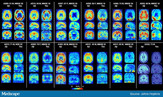
In the first article, the researchers led by Wong recruited 12 patients with Alzheimer’s disease, seven younger healthy controls, and five older healthy controls for brain PET scans. An additional six older healthy controls were recruited for full-body scanning.
The study was divided into three parts. In the first part, three designated tau tracers were tested, 11C-RO-963, 11C-RO-643, and 18F-RO-948, and 18F-RO-948 showed the best results.
In the second part of the study, researchers tested 18F-RO-948 with additional brain imaging in five patients with Alzheimer’s and five older controls, with follow-up of previously seen patients to evaluate the potential progression of tau protein tangling after an average span of approximately 16 months.
The third part of the study examined six older control patients who underwent whole-body scanning. Researchers looked at 80 different regions of the brain to evaluate how well the tracers were taken up by the brain, how well they penetrated the tissue, and how specifically they bound to the tau protein.
They found that healthy brains retained little to no tracer, whereas the brains of those with Alzheimer’s showed tau to be in regions of the brain consistent with previously reported postmortem data on filamentous tangles.
18F-RO-948 is a promising radiotracer for imaging tau pathology in Alzheimer’s disease,” the investigators write. “The tracer shows good brain uptake, has no apparent brain-penetrant radiolabeled metabolites, has a good kinetic profile, shows little or no retention in cognitively normal controls and a distribution in Alzheimer’s subjects consistent with published postmortem data.
“It is our hope that tools such as 18F-RO-948 will allow us to gain a better understanding of the pathophysiology of Alzheimer’s and, in the context of drug development, select patients for clinical trials, confirm the mechanism of action of drugs targeting pathologic tau, and monitor the effects of disease-modifying therapies regardless of whether they target tau directly,” they conclude.
In the second article, the team examined the detailed quantification of 18F-RO-948 tau binding in 11 patients with Alzheimer’s disease, five young cognitively normal controls, and five older cognitively normal controls, and verified that the compound showed reproducible results.
The study was funded by F. Hoffmann-La Roche. Wong has reported no relevant financial relationships.
J Nucl Med. 2018;59:1869-1876, 1877-1884.

After naloxone, when can opioid overdose patients be safely discharged?


Naloxone has saved thousands of lives. But can patients be safely discharged from the Emergency Department (ED) just an hour after they receive the medication that curtails drug overdoses?
According to the St. Paul’s Early Discharge Rule developed in 2000, that’s how long providers should observe patients after naloxone treatment, so long as their vital signs meet specific criteria and they are ambulatory.
But the rule was never externally validated or assessed in light of the changes that have occurred in recent years with opioid use disorder.
That’s why University at Buffalo researchers conducted the current study, published today in Academic Emergency Medicine, and the first to clinically assess the rule developed at St. Paul’s Hospital in Vancouver.
Dramatic changes
“The landscape of opioid use disorder has changed dramatically,” said Brian Clemency, DO, lead author on the paper, associate professor of emergency medicine in the Jacobs School of Medicine and Biomedical Sciences at UB and an attending physician specializing in emergency medicine at Erie County Medical Center. He also is a physician with UBMD Emergency Medicine.
In 2000, he explained, naloxone was almost exclusively administered intravenously by doctors, nurses and paramedics. Today, the medication is far more widely available, including to members of the public, and is often given in the form of a nasal spray. In addition, the use of heroin and synthetic opioids, such as fentanyl and carfentanil, has increased tremendously.
“Recommendations for patient observation after naloxone administration are inconsistent,” said Clemency. “Patients can be observed for six or more hours or they can be immediately discharged with no further evaluation.
“The question is, which of these patients needs to be watched longer?” he asked. “Right now, there isn’t a really good rule. This has wide-ranging negative implications for emergency care and opioid use disorder treatment.
“It is our hope that these findings will lead to a reduction in practice variation and allow for better use of resources in the ED, while ensuring patient safety,” he added.
Tracking patients in the ED
To determine if the one-hour early discharge rule is valid, given the changes in opioid use disorder, Clemency and his colleagues launched an ambitious study at Buffalo’s Erie County Medical Center, (ECMC) a busy, urban teaching hospital affiliated with the Jacobs School.
Patients who arrived at the medical center by ambulance after receiving naloxone for suspected opioid overdose had to be enrolled and evaluated within 30-40 minutes of arrival.
One hour after receiving naloxone in the community, patients’ vital signs were evaluated, ranging from body temperature and heart rate to blood pressure and the blood oxygen level.
A total of 538 patients were included in the study. Patients were typically observed for at least four hours before being discharged.
Patients were tracked through their hospitalization for any adverse events. Medical examiner records were then reviewed for subsequent fatalities.
The authors reported that most adverse events seen in patients with normal examinations after receiving naloxone were minor and unlikely to be life-threatening.
“This rule is a way to predict which patients will have adverse outcomes after they overdose on opiates,” concluded Clemency. “The rule is simple to follow and can be used by health care providers with varying levels of training and experience.
“We anticipate this study will lead to nationally standardized recommendations for the observation of patients following the administration of naloxone for suspected opioid overdose,” he said.
Story Source:
Materials provided by University at Buffalo. Original written by Ellen Goldbaum. Note: Content may be edited for style and length.

Journal Reference:
  1. Brian M. Clemency, William Eggleston, Evan W. Shaw, Michael Cheung, Nicholas S. Pokoj, Michael A. Manka, Donald J. Giordano, Laura Serafin, Han Yu, Heather A. Lindstrom, David Hostler. Hospital Observation Upon Reversal (HOUR) With Naloxone: A Prospective Clinical Prediction Rule Validation StudyAcademic Emergency Medicine, 2018; DOI: 10.1111/acem.13567

How lifestyle drives ER-positive breast cancer


Poor diet and lack of exercise are associated with cancer development, but the underlying biology is not well understood. Advanced glycation end products (AGEs) could offer a biological link to help us understand how certain lifestyle behaviors increase cancer risk or lessen the likelihood that an anti-cancer therapy will be effective.
AGE accumulation is the natural and unavoidable result of the breakdown of nutrients, sugars and fats. AGE levels, however, can be increased by the consumption of processed foods high in sugar and fat. Certain cooking techniques, such as grilling, searing and frying, also increase AGE formation.
High AGE levels could prevent patients with estrogen receptor (ER)-positive breast cancer from responding to tamoxifen therapy, suggest preclinical findings reported by researchers at the Medical University of South Carolina (MUSC) in a recent issue of Breast Cancer Research and Treatment. The MUSC team was led by David P. Turner, Ph.D., an assistant professor in the MUSC College of Medicine and a member of the Hollings Cancer Center, who is one of the two corresponding authors on the article. Marvella E. Ford, Ph.D., a professor in the MUSC College of Medicine and associate director of Cancer Disparities at Hollings Cancer Center, is the other corresponding author.
“By showing that AGEs in the diet may impact how well breast cancer patients respond to therapy we can make breast cancer patients aware of their existence,” says Turner. “And we can design lifestyle interventions aimed at reducing AGE intake.”
AGEs cause an imbalance between molecules called free radicals and antioxidants, leading to chronic inflammation that can promote the development of a variety of chronic diseases. Furthermore, as AGEs accumulate in our organs, they cause damage that is associated with diseases such as diabetes, Alzheimer’s, cardiovascular disease, arthritis and cancer. However, AGEs have not been studied in depth in the context of cancer.
The publication by Turner, Ford and colleagues shows that elevated AGE levels lead to continual activation of pathways that promote cancer cell growth. A key molecule turned on by those pathways is important in the context of ER-positive and -negative breast cancer. This led the MUSC team to explore how AGE might affect cancer cell signaling in ER-positive breast cancer.
The MUSC team found that AGEs actually increase the phosphorylation (a process that turns on a biological pathway) of a protein called estrogen receptor alpha in a breast cancer cell line model. Adding tamoxifen to the cancer cells prevented their growth. However, adding AGEs caused them to grow once again. This could mean that patients with high AGEs are less likely to respond to tamoxifen treatment.
Turner’s team also found that a defined lifestyle intervention of exercise and dietary counseling lowered systemic levels of AGEs in overweight women with non-metastatic ER-positive breast cancer.
Next steps are to expand the published study to determine the effects of the intervention on a larger scale, while also further exploring the biological pathways in animal models. Together, they should shed light on how lifestyle interventions can beneficially affect cancer treatments by reducing AGE levels.
Story Source:
Materials provided by Medical University of South CarolinaNote: Content may be edited for style and length.

Journal Reference:
  1. Katherine R. Walter, Marvella E. Ford, Mathew J. Gregoski, Rita M. Kramer, Kendrea D. Knight, Laura Spruill, Lourdes M. Nogueira, Bradley A. Krisanits, Van Phan, Amanda C. La Rue, Michael B. Lilly, Stefan Ambs, King Chan, Tonya F. Turner, Heidi Varner, Shweta Singh, Jaime Uribarri, Elizabeth Garrett-Mayer, Kent E. Armeson, Ebony J. Hilton, Mark J. Clair, Marian H. Taylor, Andrea M. Abbott, Victoria J. Findlay, Lindsay L. Peterson, Gayenell Magwood, David P. Turner. Advanced glycation end products are elevated in estrogen receptor-positive breast cancer patients, alter response to therapy, and can be targeted by lifestyle interventionBreast Cancer Research and Treatment, 2018; DOI: 10.1007/s10549-018-4992-7

Daily Aspirin May Reduce COPD Exacerbations


Taking a daily aspirin was linked to reduced COPD exacerbations, less shortness of breath, and better quality of life, an analysis of the ongoing SPIROMICS study found.
At 3 years follow-up, aspirin users were less likely to have acute COPD exacerbations (adjusted incidence rate ratio [IRR] 0.78, 95% CI 0.65-0.94) compared with non-users, with a similar effect seen for moderate acute COPD exacerbations (IRR 0.86, 95% CI 0.63-1.18), according to Ashraf Fawzy, MD, of Johns Hopkins University in Baltimore, and colleagues.
The association was strongest among study participants reporting symptoms of chronic bronchitis at enrollment, as reported in the journal CHEST.
Aspirin use has been associated with reduced mortality in previous studies, but the newly published investigation is among the first to examine the impact of daily aspirin therapy on respiratory morbidity in COPD.
Preliminary data from this study were presented earlier this year at the American Thoracic Society annual conference.
“The reduced incidence of total and moderate acute COPD exacerbations among aspirin users was independent of concurrent respiratory or cardiovascular medication use and robust when analyzing the entire follow-up period, limiting the analysis to the first year of follow-up, and across all sensitivity analyses,” the authors wrote.
Aspirin use was also associated with lower total scores on the St. George Respiratory Questionnaire (β -2.2, 95% CI -4.1 to -0.4); reduced odds of moderate-to-severe dyspnea, score ≥2 on the modified Medical Research Council Questionnaire (adjusted OR 0.69, 95% CI 0.51-0.93); and lower COPD Assessment Test scores (β -1.1, 95% CI -1.9 to -0.2).
No difference was observed between groups in 6-minute walk distance (β 0.7 meters, 95% CI -14.3 to 15.6).
The analysis included COPD patients in SPIROMICS who self-reported daily aspirin use at study entry, 45% of the 1,698 study participants. Acute exacerbations of COPD were prospectively determined through quarterly structured telephone questionnaires for up to 3 years and categorized as moderate (symptoms treated with antibiotics or oral corticosteroids) or severe (requiring an emergency department visit or hospitalization).
Aspirin users were matched 1:1 with non-users based on propensity score, which resulted in 503 participant-pairs. The association of aspirin use with total, moderate, and severe acute COPD exacerbation was investigated using zero-inflated negative binomial models. Linear or logistic regression were used to investigate the association with baseline respiratory symptoms, quality of life, and exercise tolerance.
The researchers noted that aspirin has several systemic and local pulmonary mechanisms of action that could explain the findings, including “inactivation of platelets and reduced inflammation,” among them.
“A urinary metabolite of thromboxane A2, which is secreted by activated platelets, has been shown to be elevated among patients with COPD and represents the pathway irreversibly blocked by aspirin. Persistent systemic elevation of inflammatory markers interleukin-6 and CRP may represent a systemic inflammatory phenotype of COPD which are attenuated by aspirin in other patient populations,” the researchers wrote.
They further noted that in a 2017 study, treatment with aspirin was found to reduce pro-inflammatory cytokines in bronchoalveolar lavage samples of 33 healthy volunteers.
Study limitations cited by the researchers included the study participants’ self-reporting of daily aspirin use, without dosing information. Information on duration of aspirin use and adherence to therapy before and during the study was also unavailable. COPD exacerbations were not confirmed with medical records, which may have led to misclassification of events.
And despite propensity score matching and other efforts to avoid confounding, the researchers acknowledged that they may not have controlled for all factors that could have impacted their outcomes.
They concluded that a randomized study is needed to determine whether daily aspirin use is protective against COPD exacerbations. “Prospective randomized clinical trials of aspirin use are warranted to explore its potential effect in reducing COPD morbidity,” they wrote.
Funding for this research was provided by the National Heart, Lung, and Blood Institute, the National Institute for Environmental Health Sciences, and by the drugmakers AstraZeneca/Medimmune, GlaxoSmithKline, and others through the Foundation for the NIH and the COPD Foundation.
LAST UPDATED 

Seizure Patients Often See Swift Return to Hospital


Nearly 11% of U.S. patients hospitalized with seizures were readmitted within 30 days — most commonly for convulsions or epilepsy — a retrospective cohort study showed.
While readmitted patients were more likely to have multiple medical comorbidities, inpatient adverse events also were significantly associated with 30-day readmission, reported Leah Blank, MD, MPH, of the University of Pennsylvania in Philadelphia, and co-authors in Neurology.
This study is the first to describe a nationally representative 30-day readmission rate for seizure discharge, Blank said. “This is important because hospital readmissions are increasingly being used to assess hospital — and, perhaps in the future, provider — performance,” she told MedPage Today.
Medicare payments are already docked for centers with poor performance on 30-day readmissions after an initial admission for a number of diagnoses, including heart attack, she added. “This list of penalized diagnoses is likely to expand and we think seizure is likely to be the first neurologic disease to be included.”
Readmission metrics are monitored as markers of quality and cost-effective care, noted Nathalie Jette, MD, of the Icahn School of Medicine at Mount Sinai in New York, and colleagues in an accompanying editorial.
“Current health care regulations allocate monetary incentives directed at enhancing quality of care that is also cost saving,” they wrote. “Although epilepsy is not currently within the list of diseases being penalized for readmission within 30 days by the Centers for Medicare & Medicaid Services, this will undoubtedly change as it is considered by many an ambulatory care sensitive condition.”
On a national population-based level, little was known regarding risk factors for readmission after seizure-related discharges, Jette and co-authors added.
In this study, Blank and her group studied emergently hospitalized adults with a primary discharge diagnosis of seizure or epilepsy, sampled from the Healthcare Cost and Utilization Project’s 2014 Nationwide Readmissions Database. They excluded elective hospitalizations, hospitalizations in which the patient left against medical advice, or hospitalizations occurring near the end of the year due to insufficient follow-up.
The researchers identified 139,800 hospitalizations that met inclusion criteria for 30-day readmission. Of these, 15,094 patients (10.8%) were readmitted within 30 days. The most common primary reasons for readmission were epilepsy or convulsions (17%) and sepsis (7%).
In multivariate logistic regression models, several variables were tied to 30-day readmission:
  • Higher Elixhauser comorbidity score (OR 2.28 for 4+ vs ≤1 condition, 95% CI 2.01-2.58)
  • Longer index admission stay (OR 1.78 for 7+ days, 95% CI 1.63-1.95)
  • Public insurance (compared with private insurance: OR 1.48 for Medicare, 95% CI 1.34-1.62, or OR 1.39 for Medicaid 95% CI 1.26-1.54)
  • Discharge to an inpatient care facility (OR 1.32 vs with discharge home, 95% CI 1.23-1.42)
  • Documented adverse events during the index admission (OR 1.17, 95% CI 1.06-1.30)
When adverse events were analyzed in more detail, medication adverse events no longer were statistically significant (OR 1.12, 95% CI 0.99-1.26), but medical or surgical events were (OR 1.34, 95% CI 1.10-1.62).
“The most common reason for readmission was epilepsy or convulsion, which suggests that there is an opportunity for improvement as patients are not being readmitted for unforeseeable complications,” Blank said.
Adverse medical events need to be examined on granular level to better understand the health care structure — neurology ICU, continuous EEG, and neurology nursing, for example — and the processes and protocols they are associated with, she added: “Although documented adverse events were uncommon, readmitted patients were more likely to have had an adverse event during their original admission, which again suggests we can continue to improve the care that we deliver to seizure patients.”
Outpatient data was not available in this dataset, so the researchers could not study relationships between outpatient follow-up and readmission. They also could not assess epilepsy severity or information about social support, presence of a care partner, marital status, education, or anti-seizure medication adherence.
The research was supported by the Mirowski Family Fund and by the American Epilepsy Society/Epilepsy Foundation Research and Training Fellowship for Clinicians and a Neurologic Clinical Epidemiology Training Grant.
The researchers reported no conflicts of interest.
Editorialists reported relationships with Alberta Health, Eisai, Medtronics, Sunovion Pharmaceuticals, Epilepsy Study Consortium, GW Pharmaceuticals, NeuroPace, Novartis, Supernus, Upsher-Smith Laboratories, UCB Pharma, and Vivus Pharmaceuticals.

Lutikizumab Ineffective for Erosive Hand Osteoarthritis


Target Audience and Goal Statement:
Rheumatologists, internists, and family medicine specialists
The goals were to measure the efficacy and safety of lutikizumab, an anti-interleukin-1α (IL-1α) and anti-interleukin 1β (IL-1β) dual variable domain immunoglobulin in patients with erosive hand osteoarthritis (OA).
Questions Addressed:
  • Did this drug relieve pain in patients with erosive hand OA?
  • What are the safety, pharmacokinetics, and pharmacodynamics of lutikizumab in these patients?
  • Did this treatment improve other symptomatic, functional, and structural endpoints for this drug compared to placebo?
Study Synopsis and Perspective:
In a phase IIa study in 110 patients with erosive hand OA, lutikizumab, a dual variable-domain immunoglobulin that inhibits IL-1α and IL-1β, was no more effective than placebo in reducing pain and inflammation.
Hand OA is common, particularly among older women, and is associated with substantial disability, including pain and swelling, but available treatments are limited, and no disease-modifying agents have demonstrated efficacy to date.
Preclinical studies and mouse models indicated that IL-α and IL-β were potential mediators of synovitis, cartilage damage, and bone loss in patients with this condition, but this clinical trial did not confirm earlier research regarding pain, nor did it improve other symptomatic, functional, and structural secondary endpoints.
For this randomized placebo-controlled study Kloppenburg and colleagues enrolled 131 adults with active inflammation of the hand joints, with pain ratings of six or higher on an 11-point scale, and at least one erosion of an interphalangeal joint. All underwent radiography and MRI of the hand joints at baseline and week 26. Radiographs were scored according to the Osteoarthritis Research Society International (OARSI) criteria and MRIs according to the Outcome Measures in Rheumatology/Hand Osteoarthritis MRI Scoring system (OMERACT/HOAMRIS).
Participants’ mean age was 66, and most were white women. Mean duration of OA was 11 years, mean Australian/Canadian Osteoarthritis Hand Index (AUSCAN) pain score at baseline was 39 (pain score ranges from 0 – 50), and mean tender and swollen joint counts were 12 and 6, respectively.
Patients were randomized to receive subcutaneous lutikizumab, 200 mg every 2 weeks for 24 weeks, or placebo subcutaneous injection. They were permitted rescue medication for pain with acetaminophen or ibuprofen.
At week 16, change from baseline in pain as measured on the AUSCAN was similar for patients who had been randomized to lutikizumab (−9.2, 95% CI −13.8 to −4.6) or placebo (−10.7, 95% CI −15.4 to −6), according to Margreet Kloppenburg, MD, PhD, of Leiden University Medical Center in the Netherlands, and colleagues.
At week 16, least squares mean difference between lutikizumab and placebo in change of pain was a nonsignificant 1.5 (95% CI −1.9 to 5), the researchers reported online in Annals of the Rheumatic Diseases.
As with the pain endpoint, no significant differences were seen between the two groups in change from baseline in AUSCAN function at week 16, which was −14.6 (95% CI −22.1 to −7.1) for lutikizumab, and −17.2 (95% CI −24.9 to −9.4) for placebo, for a mean difference of 2.5 (95% CI −3.2 to 8.3).
Other secondary endpoints also had similar results for lutikizumab and placebo at week 26, respectively:
  • Tender joints, −5.8 vs −4.7, P=0.32
  • Swollen joints, −2.2 vs −1.8, P=0.64
  • OARSI joint space narrowing, 0.03 vs 0.14, P=0.51
  • HOAMRIS synovitis, 0.85 vs 0.92, P=0.89
  • Joints with synovitis on MRI, 0.54 vs 0.46, P=0.79
Source Reference: Annals of the Rheumatic DiseasesDec. 14, 2018 DOI: 10.1136/annrheumdis-2018-213336
Study Highlights: Explanation of Findings
Erosive hand OA is an unmet medical need: there are no treatments currently which prevent structural damage, although non-steroidal anti-inflammatories (NSAIDs), topical and oral, may relieve pain. In a previous study with an inhibitor of another pro-inflammatory cytokine, antitumor necrosis factor (anti-TNF), demonstrated that this approach prevented structural damage though it did not produce pain relief. An antimalarial drug, hydroxychloroquine, was also tested in a randomized placebo-controlled study known as HERO (Hydroxychloroquine Effectiveness in Reducing symptoms of hand Osteoarthritis), but also produced no significant symptomatic, or radiographic, efficacy in patients with severe hand OA.
Preclinical studies with IL-1α and IL-1 β blockade, however, indicated that inflammatory cytokines IL-1α and IL-1β are involved in the pathogenesis of OA. “IL-1α and IL-1β bind to the IL-1 type 1 receptor, leading to the production of proinflammatory molecules, proteases and other mediators, which result in joint pain, inflammation and cartilage destruction,” the researchers said.
In a mouse model of human OA, inhibiting or blocking the two interleukins resulted in a decline in OA progression, and in a phase I trial of knee OA the treatment it was associated with decreases in neutrophils, C-reactive protein (CRP) and biomarkers of synovitis, such as matrix metalloproteinase-degraded collagen types I and III.
Although the drug treatment did lead to significant declines in serum levels of IL-1α and IL-1β and in levels of neutrophils, CRP levels, and matrix metalloproteinase-degraded collagen type 1, it did not resolve clinical signs and symptoms of erosive hand OA in this study. The pharmacokinetics of preclinical and phase 1 studies were replicated and confirmed in this trial, but that did not affect symptoms.
Adverse events and serious adverse events were similar in the two groups, but more patients in the lutikizumab group reported injection site reactions. Study withdrawals, because of adverse events, occurred in five patients receiving lutikizumab and two given placebo, and two patients receiving the active treatment withdrew from the study because of neutropenia. Low-density lipoprotein cholesterol levels increased more in the active treatment group.
One strength of the study was its stringent enrollment of only those with moderate to severe inflammation.
An important limitation of the study is the possibility that 26 weeks was not long enough to detect changes in pain, function, and structure, researchers noted. In addition, researchers did not measure hand joint synovial fluid levels of the drug or the target cytokines, which leaves open the question of whether the inflammatory cytokines had been reduced locally in the joint, which could impact pain and function. They also noted that they had no adequate explanation for the low use of non-steroidal anti-inflammatories (NSAIDS) or other pain relievers, given the high average level of pain patients had at baseline.
“In conclusion, despite adequate systemic lutikizumab pharmacodynamic effects, lutikizumab did not significantly improve clinical outcomes or imaging outcomes in patients with erosive hand OA compared with placebo, suggesting that targeting IL-1 may be ineffective for the treatment of erosive hand OA,” the authors wrote.
Nancy Walsh wrote the original study for MedPage Today

Youth Opioid Deaths Nearly Tripled Over Past Two Decades


Nearly 9,000 youth died from opioid poisonings, with the mortality rate increasing nearly threefold in the past two decades, according to a retrospective study.
The overall pediatric mortality rate from prescription and illicit opioid poisonings rose 268.2% — increasing from 0.22 per 100,000 (95% 0.19-0.25) in 1999 to 0.81 per 100,000 (95% CI 0.76-0.88) in 2016 (P for time effect<0.001), reported Julie Gaither, PhD, MPH, RN, of Yale School of Medicine in New Haven, Connecticut, and colleagues.
Opioid-related deaths were most common amongst teenagers, and the mortality rate for adolescents ages 15-19 increased by 252.6% across the study period, they wrote in JAMA Network Open.
However, all age groups experienced a rise in rates over time, with mortality rates among the youngest children ages 0-4 rising by 225.0%, the authors noted.
“The primary thing is that we need to consider — public health officials, legislators, clinicians, and parents — is how everyone is affected in the home when an adult brings an opioid into the house,” Gaither told MedPage Today, adding that this includes administering safety recommendations and disposal instructions based on who is in the home along with opioid prescriptions.
Although differing in magnitude, pediatric mortality rates followed a similar temporal and drug use pattern as adult opioid-related mortality rates, with pediatric mortality rates demonstrating a steady linear increase until roughly 2008, the authors reported.
Gaither said that similar to adult trends, after a brief plateau from 2012 to 2014, youth mortality rates rose again starting around 2014, when regulations tightened around opioid prescriptions, and a surge in synthetic opioid deaths occurred.
Although prescription opioids are primarily driving the epidemic, the introduction of drugs like illicitly manufactured fentanyl likely contributes to an increased number of children who are exposed, with nearly one third of teen deaths in this study attributed to synthetic opioids since 2014, Gaither noted.
“With our data, we were not able to distinguish between what was pharmaceutical fentanyl and what was illicitly manufactured fentanyl, and no study is able to do that because you cant look at that, but these are likely illicitly manufactured drugs,” Gaither said. “That’s very troubling that teens are getting hold of these illegal drugs that are 50 to 100 times more potent than heroin.”
In this study, researchers analyzed cross-sectional data from the Centers for Disease Control and Prevention (CDC) Wide-Ranging Online Data for Epidemiologic Research (WONDER) database to determine the number of poisonings from prescription and illicit opioids across the study period. Deaths were identified using ICD-10 codes.
Overall, 8,986 youth died from opioid poisonings across the study period, with 7,921 (88.1%) deaths involving teenagers ages 15-19, 605 (6.7%) involving children ages 0-4 and 364 (4.1%) involving children ages 10-14. The majority of deaths occurred in non-Hispanic white children (79.9%) and in boys (73.1%), the researchers reported.
In teens ages 15-19, the authors found that heroin was responsible for 1 in 4 deaths (23.6%) and from 1999 to 2016, the rate of fatal heroin overdoses increased by 404.8%, while the rate of prescription opioid deaths in this age group increased by 94.7%. Additionally, synthetic opioids resulted in an increased mortality rate of 2,925% within this age group.
Most deaths in this study were unintentional, with just 5% attributed to suicide and 2.4% to homicide. Gaither said further research should investigate the circumstances in which these deaths occur, and if parental opioid abuse is contributing to them.
Additionally, many deaths took place outside of a medical setting, with 38% of youth dying at home. Just 10.4% of deaths occurred in inpatient settings and 24.1% in emergency departments. Gaither said this suggests these deaths likely occur suddenly and take place before emergency services can reach a child who has been exposed to opioids.
Because this study relies on data from death certificates, it is possible that some deaths are misclassified in terms of the cause and manner of death, the authors reported. Researchers also noted that this study is limited because they were not able to determine the circumstances of death, as well as whether deaths involved pharmaceutical or illicitly manufactured fentanyl. It’s possible, therefore, that the number of deaths found across the study period could be underestimated, they reported.
This study was supported by the National Center for Advancing Translational Sciences, National Institutes of Health.
Gaither disclosed grants from the NIH for this study.